Download
1 / 28
280 likes | 297 Vues
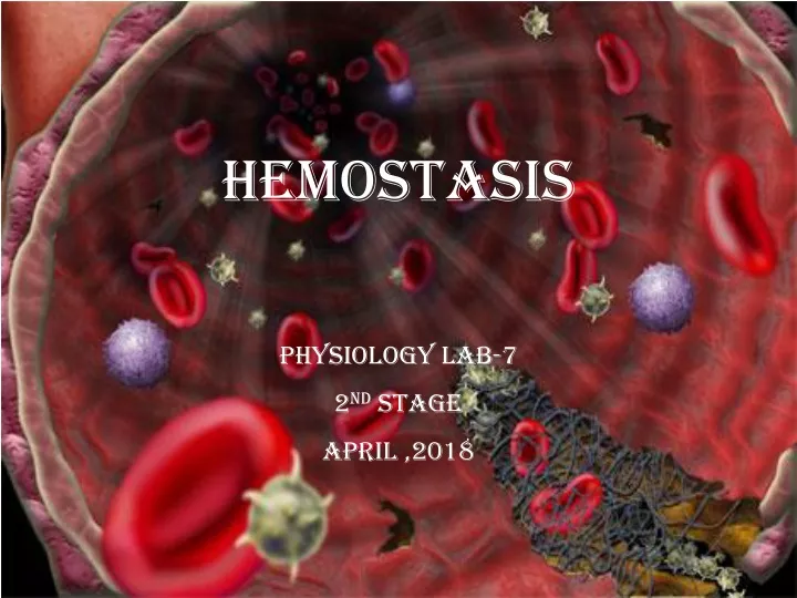
hemostasis Physiology Lab-7 2 nd Stage April ,2018. HEMOSTASIS. DEFINITION - Heme = blood - stasis = to stop

E N D
hemostasis Physiology Lab-7 2nd Stage April ,2018
HEMOSTASIS • DEFINITION - Heme = blood - stasis = to stop • It is the process of forming clots in the wall of damaged blood vessels & preventing blood loss while maintaining blood in a fluid state with in the vascular system. • Defects in hemostasis can lead to an increased risk of bleeding (hemorrhage) or clotting (thrombosis).
Stages of Hemostasis Vascular Constriction Formation of Platelet Plug Formation of blood clot Fibrinolysis
Events in Hemostasis • Vascular Constriction -Damaged blood vessels constrict • Formation of platelet Plug - Platelets adhere to damaged endothelium to form platelet plug (primary hemostasis). • Blood Coagulation - Clots form upon the conversion of fibrinogen to Fibrin, and its addition to the platelet plug (secondary hemostasis).
2- STAGES OF PRIMARY HEMOSTASIS Platelet Adhesion Platelet Activation Platelet Aggregation
3- Secondary hemostasis If there is a large hole in the blood vessel, a blood clot is additionally required. Cascadeof reactions It states that ‘inactive’ enzymes are activated, and the ‘activated’ enzymes in turn activates other inactive enzymes until final step is reached.
THE CLOTTING MECHANISM INTRINSIC EXTRINSC Collagen Tissue Thromboplastin XII XI VII IX VIII X V FIBRINOGEN (I) PROTHROMBINTHROMBIN (III) (II) FIBRIN
4-FIBRINOLYTIC PHASE The fibrinolytic system does not allow the fibrin clot to grow and block a vessel, which would cause serious complications. The dissolution of a clot, called fibrinolysis (dissolving of fibrin fibers), is brought about by the formation of the active enzyme plasmin from plasminogen
HEMOSTASIS • DEPENDENT UPON: • Vessel Wall Integrity • Adequate Numbers of Platelets • Proper Functioning Platelets • Adequate Levels of Clotting Factors • Proper Function of Fibrinolytic Pathway
So What Causes Bleeding Disorders? • VESSEL DEFECTS • PLATELET DISORDERS • FACTOR DEFICIENCIES
METHOD OF STUDY • HEMOSTATIC FUNCTION TESTS -Bleeding time -Clotting time -Prothrombin time -Partial prothrombin time -Thrombin time
Note • The BT and CT are two simple tests that are used as a routine before every minor and major surgery (e.g. tooth extraction), biopsy procedures, and before and during anticoagulant therapy, whether or not there is a history of bleeding.
1-BLEEDING TIME (B.T) • Definition: • is the time interval between the skin puncture and spontaneous, unassisted (i.e. without pressure) stoppage of bleeding. The BT test is an in vitro test of platelet function. • Purpose: to detect qualitative defects of platelets. • Normal bleeding time ; 1 – 5 min.
Bleeding Time • Medical applications: The prolongation of bleeding time may be due to: • Defects in the blood vessels • Decrease number of platelets(thromocytopenia) • Defect in the function of the platelets caused by:- • drugs (aspirin, NSAIDs, Anti coagulants, Sulfonamides, Diuretics,etc.) • Inherited diseases (VON WILLEBRAND’S DISEASE.
Materials and methods Bleeding Time • Lancet • Stop watch • Circular filter paper • Alcohol
Bleeding Time Materials and methods • A disposable lancet is used to make cut into the finger usually. • A stopwatch is started immediately and every 30 seconds filter paper is used to draw off the blood. • The time from when the incision is made until all bleeding has stopped is called the bleeding time. • Note:The filter paper should not touch the edge of the clot as this may disturb the formation of the platelet plug. • The test is finished when bleeding has stopped completely. • Count the number of blood spots and express your result in minutes and seconds.
2-PROTHROMBIN TIME • Measures Effectiveness of the Extrinsic Pathway. • Normal ratio 0.9-1.2
3- PARTIAL THROMBOPLASTIN TIME Measures Effectiveness of the Intrinsic Pathway and common pathway NORMAL VALUE 25-35 SECS
4-Thrombin Time (TT) • Time to clot formation after addition of thrombin to citrated blood • Normal value : less than 15 seconds
5-CLOTTING TIME ( C.T )(COAGULATION TIME) • Definition : is the time interval between the entry of blood into the glass capillary tube, or a syringe, and formation of fibrin threads • Normal Clotting Time :3 – 6 min. • Prolonged clotting time is due to severe deficiency of any of the coagulation proteins. • Weak friable clot called hypofibrinogenaemia. • Method : capillary tube method.
Clotting time - capillary method • Material • Sterile disposable pricking lancet. • Stop watch • Dry capillary tube (non heparinized) • Cotton Swab . • 70 % ethyl alcohol
Clotting time - capillary method • Clean your finger with alcohol • Prick the finger by a lancet and note the time using a stop watch • Load a capillary tube to at least ½ full • After about 2 mins, take the loaded capillary tube between your thumb and forefingers and gently break in half • Slowly pull the ends part to see the insoluble fibrin strands • Do a break every 30 sec, once the clot is formed we record the time
The clotting of blood with this method involves both the intrinsic and the extrinsic systems of clotting. There is injury to the blood (coming in contact with glass, intrinsic pathway), and the injury to the tissues (extrinsic pathway).
The coagulation time is increased in the following conditions: 1. Hemophilias 2. von Willebrand disease. 3. Afibrinogenemia and dysfibrinogenemia 4. Vitamin K deficiency it acts a cofactor in the synthesis of prothrombin, and factors VII, IX and X 5. Liver diseases 6. Anticoagulant therapy. Patients receiving heparin or warfarin show an increased CT. 7 .Newborns. Newborns, especially premature babies sometimes have a tendency to bleed because the plasma levels of certain factors are low