Download
1 / 39
410 likes | 443 Vues
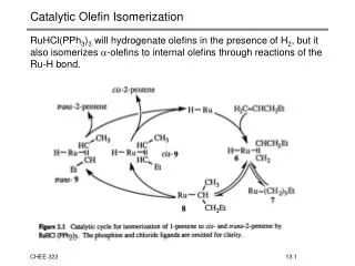
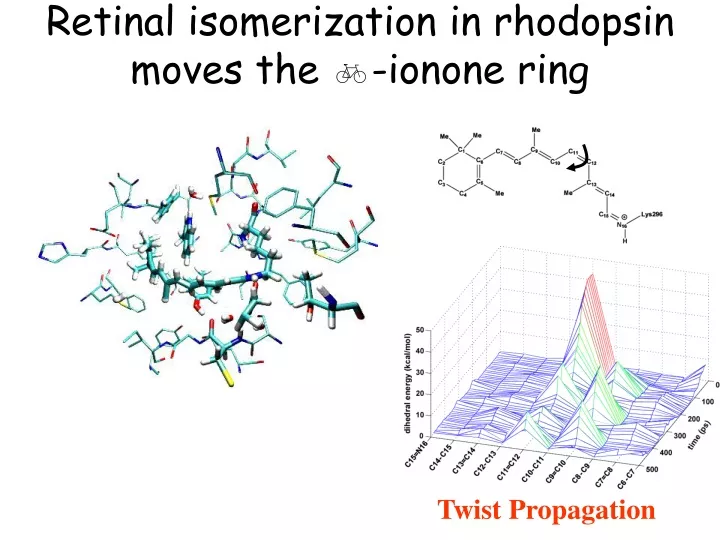
Retinal isomerization in rhodopsin moves the b -ionone ring. Twist Propagation. How does photon energy is stored in rhodopsin?. S 1. S 0. Internal (bonded) and interaction energy of retinal with its surrounding. Isomerization of retinal allows helix VI to rotate freely.

E N D
Retinal isomerization in rhodopsin moves the b-ionone ring Twist Propagation
How does photon energy is stored in rhodopsin? S1 S0 Internal (bonded) and interaction energy of retinal with its surrounding
Isomerization of retinal allows helix VI to rotate freely Torque applied to rotate helix VI before (red) and 5 ns after isomerization (blue)
Major conformational Changes in 10 ns Helix Movement Cytoplasmic Changes
Color Vision cone cells N hn N all-trans 13-cis Visual receptors of rhodopsin family are classified based on their color sensitivity
Comparison of color visual receptors and rhodopsin Each of the human cone receptors is only 40% identical with human rhodopsin. The human blue color receptor is only 40% identical with the human green and red receptors. Only 15 out of 364 residues of human green and red cone receptors are different.
Green to red Green Red Ala180 Serine Phe277 Tyrosine Ala285 Threonine Electrostatic interaction of retinal and the binding pocket, in favor of excited state or in disfavor of the ground state. Too bad we do not have the structures, but we could use Rh.
VMD examination of rhodopsin and color visual receptors Sequence alignment of color receptors
Color vision in other mammals Sequences are all available and one could mutate them in rhodopsin, and try to explain the effect of amino acid exchange on the spectral properties.
Physics of Color Deficiency Normal Males are afflicted more often than females! Deutan (M-cone)
Signal transduction over a 5 nm distance Retinal-rhodopsin complex Amphiphilic H8 Trimeric G-protein (Transducin)
Photoinduced isomerization = Ligand binding hn Neurotransmitters Hormones Drugs … G-protein activation G-protein activation Ligand induced conformational changes is the main mechanism of activation of GPCRs
Olfaction The organization and components are very similar to vision. So we expect similar receptor architecture for olfaction.
Olfactory Receptors extracellular Red: variable Blue: conserved cytoplasmic A few hundred types of ORs are present in human beings. Each neuron has only one type of OR.
Cell Signaling of Olfaction cAMP Signaling in vision cGMP
Ligands for Olfactory Receptors Small molecules – Stereoselecitve binding
Olfactory Receptor Genes Fraction of OR pseudogenes in different species Rat gene: more than 1000 ORs All functional Human genome: 500-750 Ors (One of the largest gene family in human beings) More than half are pseudogenes
Single cells too can smell and see! The response of bacterium is moving toward or away from the signal chemotaxis phototaxis
Organization of Flagella Proton motive force – not ATP
Chemotaxis and Phototaxis V = 25 mm/s ccw cw ccw 2 mm chaotic A motilebacterium can move 10 times its body length in one second.
Effective motion of the bacterium in the absence of any signal: R A N D O M W A L K
Random walk Chemoattractant Biassed random walk Chemorepellent Biassed random walk
Animations: Chemotaxis
Archaeal Sensory Rhodopsins bR sRI signal energy sRII hR Prof. Carl Woese (UIUC) wins the Crafoord Prize in Biosciences http://www.news.uiuc.edu/news/03/0213crafoord.html • Bacteriorhodopsin (bR) • Halorhodopsin (hR) • Sensory rhodopsin I (sRI) • Sensory rhodopsin II (sRII)
Oxygen and light together could be very harmful for the cell In aerobic condition: electrogenic pumps are not needed (bR/hR) Attractant response is not needed (sR-I) These proteins are repressed; sR-II (repellent sensory rhodopsin) is expressed to help find the dark and avoid oxidative damage to the cell. Maximal absorption of sR-II (498 nm) matches the highest intensity wavelength of sunlight at the surface of the Earth. In anaerobic condition: bR/hR/sR-I are expressed; and sR-II is suppressed.
Bacterial Rhodopsins Single protien of PM (568nm)
Slower cycle in sR-I and sR-II sR-I: Attracted to light at > 520 nm; repelled by to UV – anoxic condition sR-II: Repelled by < 500 nm – (490nm) constitutively produced – phoborhodopsin Htr – similar to chemo-receptors
Structure of the Sensory rhodopsin II/transducer complex: a molecular basis for transmembrane signalling
Cytoplasmic view Activation is based on the interaction of Helix F and TM2 Dark color: high B factor, mobile
Inherent proton pump activity of sR-II is blocked after complex formation with HtrII
Receptor-transducer interface Structure is strikingly very similar to Np-sR-II alone; only Tyr199 is different (90 degree rotated) Interface mainly vdW, only a few H-bonds
Remarkable similarity between bR and sR-IIwhy different functions? If you decouple it from Htr-II, it functions as a proton pump!!
Similarities of conformational changes in retinal proteins Displacement of helices F and G in bR is responsible for the opening of the cytoplasmic half channel and entrance for water molecules necessary for reprotonation of retinal Schiff base.
Similarities of conformational changes in retinal proteins Rotation of helix VI (F) in Rh is one of the major conformational changes triggering the activation of transducin (G-protein).
Similarities of conformational changes in retinal proteins Outward motion of helix F in sR-II causes the rotation of one of the two helices in the transmembrane region of the transducer and its further conformational change.
Importance of protein-lipid interaction Kinetic of Rh photocycle Kinetic of bR photocycle … and probably also the kinetics of sR-II photocycle, can be influenced by the lipid composition of the membrane
Next week Membrane channels:Aquaporins