Download
1 / 13
130 likes | 428 Vues
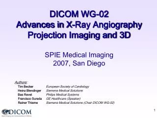
DICOM WG-02 Advances in X-Ray Angiography Projection Imaging and 3D SPIE Medical Imaging 2010, San Diego . Authors : Heinz Blendinger Siemens Healthcare Bas Revet Philips Healthcare Francisco Sureda GE Healthcare (Chair DICOM WG-02) . Presentation Outline. Introduction

E N D
DICOM WG-02 Advances in X-Ray Angiography Projection Imaging and 3D SPIE Medical Imaging 2010, San Diego Authors: Heinz Blendinger Siemens Healthcare Bas Revet Philips Healthcare Francisco Sureda GE Healthcare (Chair DICOM WG-02)
Presentation Outline Introduction • Overview of X-Ray Angiography in DICOM X-Ray N-Dimensional Presentation Cases • 3D Shutter • Volume Subtraction • Stabilized Region in Moving Volume • Trajectory Tracking in Volume • 2D-3D Blending Conclusion
Overview of X-Ray Angiography in DICOM Work in Progress Approved in the Standard X-Ray Acquisition Follow-up of IHE REM Profile Supp 94: Radiation Dose Reporting CR-DX Dose Reporting 2D Projection Images Supp 83: Enhanced XA/XRF Supp 139-LB: Enhanced XA Informative Annex Supp 140: Presentation State 3D Reconstruction X-Ray 3D Informative Annex Supp 116: X-Ray 3D Storage N-Dimensional Presentation State
Workflow in X-Ray N-Dimensional Presentation Enhanced XA Storage SOP Class X-Ray Acquisition Procedure X-Ray 3D Storage SOP Class Reconstruction Procedure In progress N-D Presentation State SOP Class Calibration Data Proprietary X-Ray Calibration Procedure Visualization Visualization X-Ray Acquisition System X-Ray 3D Reconstruction System 3D Visualization Systems
X-Ray 3D Angiography – Rotational Acquisition Optimized 3D Reconstruction Frame #5: X-ray settings 5 Geometry settings 5 Frame #4: X-ray settings 4 Geometry settings 4 Frame #3: X-ray settings 3 Geometry settings 3 Frame #2: X-ray settings 2 Geometry settings 2 Frame #1: X-ray settings 1 Geometry settings 1
X-Ray 3D Angiography – Presentation State • Needs for 3D Angiography Presentation • Presentation features common to all 3D modalities (rendering...) • Speficic presentation of X-Ray 3D Angiography: • Acquisition 3D shutter (e.g. collimation) • Volume Subtraction and voxel shift • Stabilized point in all volumes (e.g. cardiac wall motion, stent stabilized) • Catheter tracking trajectory in one volume • 2D-3D blending presentation (3D conic projection on 2D fluoroscopy) • N-Dimensional Presentation State • Work Item 2008-04-C. Addresses needs of multi-modalities • Led by Working Group 11, participation of Web3D Consortium and other DICOM working groups • Supplement in progress...
X-Ray 3D Angiography – Presentation State Punch Holes Clipping Planes Frames Volume Regions to define 3D shutter • Proposal for Volume Region Sequence: • Region Purpose : DT: “DISPLAY SHUTTER“ or “ROI“ • Region Display Mode : DT: “DISPLAY“ or “HIDE“ • Region Definition Method : Enumerated Values • ... Punch Hole: EV: “ELLIPSE“ or “POLYGON“ Plane: Orientation, Position, inside or outside Frame: EV: “ELLIPSE“ or “POLYGON“ or “BITMAP“
X-Ray 3D Angiography – Presentation State Contrast Subtracted Contrast Frames Mask Frames Volume Subtraction • Proposal for 3D Mask Subtraction Sequence: • Mask Operation: DT: “Volume Selection“ • Applicable Volume: Pair of Frame Numbers (e.g. N+1, N+k) • Mask Volume: Pair of Frame Numbers (e.g. 1, N) • Mask Voxel Shift: Triple of Float Numbers • Mask Contrast Registration 4x3 matrix • Mask Operation Explanation: Free Text • Mask Selection Mode: DT: SYSTEM or USER • Recommended Viewing Mode: DT: SUB or NAT
X-Ray 3D Angiography – Presentation State Stabilized region in moving volume Applicable to 4D data (i.e. 3D volumes with time) Goal: Stabilize the position and orientation of an object during the dynamic view • Proposal for Stabilized Region Sequence: • // contains as many items as number of volumes • Applicable Volume: Pair of Frame Numbers • Point: 3D coordinates • View Angle: 3D view angle • The result is an animated sequence to view the volumes stabilized at a given point: • a) with same volume orientation • b) or changing the orientation of the volume to keep the same orientation of the object of interest
X-Ray 3D Angiography – Presentation State Trajectory Tracking in Volume Applicable to 3D data Goal: View the volume along a pre-defined trajectory of the camera Example: • During the planning phase of a catheterization intervention • Define a 3D curve from point A to point B inside an artery (i.e. catheter trajectory) • Define an animated sequence to view the progress of the curve: • a) from the outside of the volume, with same volume orientation • b) from the outside of the volume, changing the orientation of the volume to be perpendicular to the tip of the curve • c) from inside the artery (fly-thru) as virtual endoscopy A B
2D-3D Blending in X-Ray Angiography (1/2) X-Ray 3D Reconstruction X-Ray Rotational Acquisition 3D Viewing Settings 3D View X-Ray 2D Projection SOP Class X-Ray 3D Storage SOP Class new 3D Segmentation Segmentation SOP Class Conic Projection 2D Presentation 3D Presentation Same Frame Of Reference 2D-3D Blending X-Ray 2D Projection SOP Class 2D View 2D Presentation SOP Class X-Ray Acquisition X-Ray Acquisition System
2D-3D Blending in X-Ray Angiography (2/2) 3D Image (CT, …) 3D Viewing Settings 3D View CT Storage SOP Class new 3D Segmentation Segmentation SOP Class new Conic Projection 2D Presentation 3D Presentation Registration to Isocenter System 2D-3D Blending X-Ray 2D Projection SOP Class 2D View 2D Presentation SOP Class X-Ray Acquisition X-Ray Acquisition System
Conclusion Enhanced XA (2D) • Supplement 139 in Letter Ballot. Informative (DICOM Part 17) • Will facilitate the adoption of the Enhanced XA SOP Class (Sup 83) XA Presentation State (2D) • Supplement 140 in Final Text X-Ray 3D Angiography • New IOD approved in Standard 2007 (Supplement 116) • Application cases (Informative)- Work In Progress N-D Presentation State • N-D Presentation State - Work In Progress To get involved in WG-02 developments: contact chairman at francisco.sureda@ge.com