Download
1 / 19
200 likes | 374 Vues
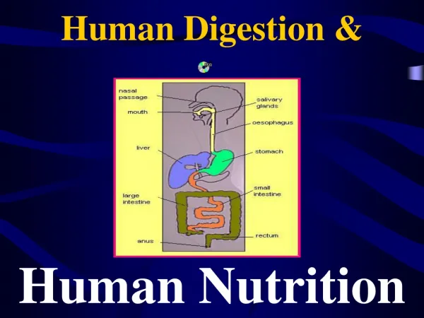
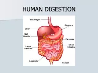
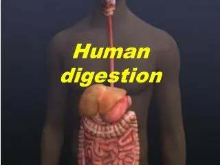
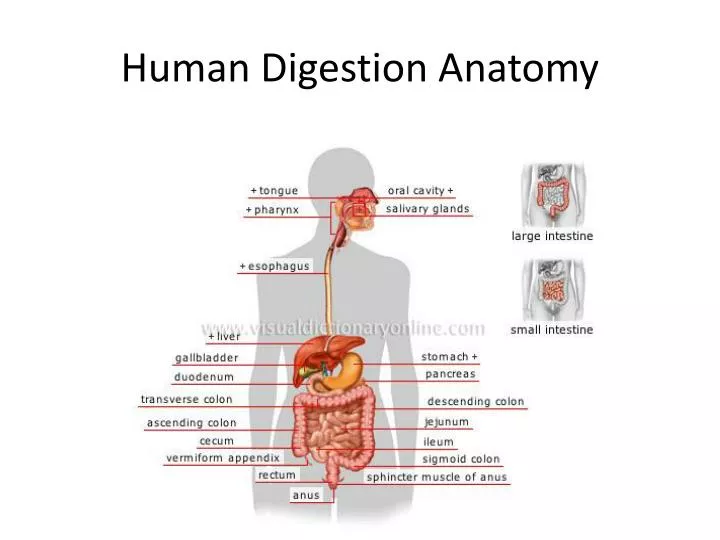
Human Digestion Anatomy. Mouth/Pharynx. Teeth c hew food into smaller pieces Tongue pushes chewed food to throat. Salivary glands. Secrete saliva to moisten food Begins the digestion of starches. Epiglottis. Flap that closes windpipe when swallowing Helps prevent choking. Trachea.

E N D
Mouth/Pharynx • Teeth chew food into smaller pieces • Tongue pushes chewed food to throat
Salivary glands • Secrete saliva to moisten food • Begins the digestion of starches
Epiglottis • Flap that closes windpipe when swallowing • Helps prevent choking
Trachea • Windpipe, not part of digestive system
Esophagus • Food tube, connects mouth to stomach
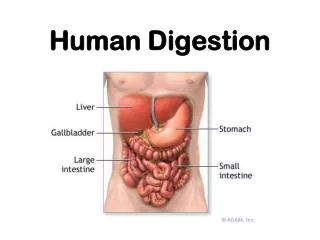
Stomach • Muscular pouch that mixes food • Digestive juices begin breakdown of proteins
Pyloric sphincter • Do-nut shaped muscle between stomach and duodenum • Controls entry of food mass into small intestines • Ground up food is called chyme
Liver • Distribution center for nutrients • Makes bile
Gall bladder • Stores bile for fat breakdown
Duodenum of small intestines • 1st 12-14 inches of small intestines • Final digestion of food occurs • Bile/fats Pancreatic juices/all food types
Pancreas • Makes enzymes that break down all type of foods
Small Intestines • Nutrient molecules absorbed into blood stream by villi and sent to liver • Ave. length 23 feet
Appendix • No real function; can become infected and removed by surgery
A possible scenario for the progression from a fully functional cecum to the current human appendix was put forth by Charles Darwin.[13] He suggested that the appendix was used for digesting leaves as primates. It may be a vestigial organ of ancient humans that has degraded to nearly nothing over the course of evolution. The cecum enables it to host bacteria that specifically help to break down cellulose. Human ancestors may have also relied upon this system when they lived on a diet rich in foliage. As people began to eat more easily digested foods, they may have become less reliant on cellulose-rich plants for energy. As the cecum became less necessary for digestion allelesbecame more frequent and the cecum continued to shrink. After millions of years, the once-necessary cecum degraded to be the appendix of today
Large Intestines • Water reabsorbed back into blood • Bacteria feed on undigested matter as it moves to rectum
Eyes, Nose, Tongue • Senses of sight , smell, and taste help to determine food choices and safety
Rectum • Stores solid waste until ready for removal
Anus • End of digestive tract; solid waste leaves body • Process of elimination is called defecation