Download
1 / 144
1.44k likes | 1.6k Vues
Chapter 8 The Integumentary System: The Protective Covering. Multimedia Directory. Slide 12 Integumentary System Exercise Slide 15 Skin Features Exercise Slide 51 Wound Repair Animation Slide 52 Decubitus Ulcers Video Slide 60 Burn Care Video Slide 73 Hair Exercise

E N D
Chapter 8 The Integumentary System: The Protective Covering
Multimedia Directory Slide 12 Integumentary System Exercise Slide 15 Skin Features Exercise Slide 51 Wound Repair Animation Slide 52 Decubitus Ulcers Video Slide 60 Burn Care Video Slide 73 Hair Exercise Slide 94 Pressure Sore Animation Slide 95 Decubitus Ulcer Video Slide 103 Eczema Video Slide 113 Skin Cancer Video Slide 133 Intradermal Drugs Video Slide 134 Subcutaneous Injections Video
Multimedia Directory (cont’d) Slide 143 Emergency Medical Technicians Video Slide 144 Nursing Video
Introduction Integumentary system protects body from environmental damage Skin forms protective barrier, shielding body from elements and pathogens, as well as performing several other vital functions Skin is essential to well-being, helps to regulate body temperature, and contains many accessory organs such as nail, hair, and glands
Learning Objectives Discuss the functions of the integumentary system List and describe the layers of the skin Explain the healing process of skin Describe the structure and growth of hair and nails
Learning Objectives (cont’d) Explain how the body regulates temperature through the integumentary system Describe various skin diseases, causative agents, and their related treatments
Pronunciation Guide Alopecia Apocrine Carotene Corium Ecchymosis Eccrine Epidermis Epithelial cells (al-oh-PEE-she-ah) (APP oh crin) (CARE eh teen) (CORE ee um) (ek ee MOH sis) (EKK rin) (ep ih DER miss) (ep ih THEE lee al) Click on the megaphone icon before each item to hear the pronunciation.
Pronunciation Guide (cont’d) Keratin Keratinization Lesion Lunula Melanin Melanocytes Pustule Sebaceous gland Seborrheic keratosis (KAIR ah tin) (KAIR ah tin eye ZAY shun) (LEE zhun) (LOO nyoo lah) (MELL an in) (mell AN oh sights) (PUS tyool) (see BAY shuss) (SEB oh REE ik KERR ah TOH sis) Click on the megaphone icon before each item to hear the pronunciation.
Pronunciation Guide (cont’d) Sebum Scabies Squamous cells Stratum corneum Subcutaneous fascia Tinea Vesicles (SEE bum) (SKAY beez) (SKWAY muss sells) (STRAY tum core NEE um) (sub cue TAY nee us FAY she ah) (TIN e ah) (VES ih koolz) Click on the megaphone icon before each item to hear the pronunciation.
System Overview Integumentary system is comprised of skin and its accessory components including hair, nails, and associated glands
System Overview (cont'd) Integumentary system performs several vital functions: Protection from pathogens Balances fluid levels Stores fatty tissue for energy supply Produces vitamin D (with help from sun) Provides sensory input Helps to regulate body temperature
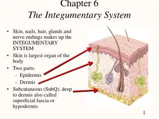
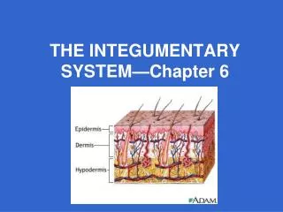
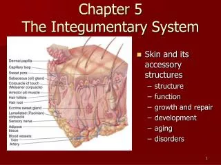
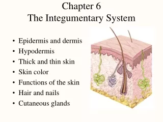
The Skin Largest organ, weighing approximately 20 pounds and covering area about 20.83 square feet on an adult Cross section of skin reveals three layers: Epidermis Dermis Subcutaneous Fascia
Epidermis Layer of skin we see on the outside; made up of five or six even smaller layers of tissue There are no blood vessels or nerve endings in this layer Cells on surface are constantly shedding, being replaced with new cells that grow and arise from deeper region called stratum basale every 2–4 weeks
Epidermis (cont'd) Outermost layer is layer of dead cells, called stratum corneum, which are flat, scaly, keratinized epithelial cells You slough off 500 million cells every day, or about 1½ pounds of dead skin a year, allowing for rapid repair in case of injuries
Integumentary System Exercise Click here to view an interactive labeling exercise on the features of the integumentary system. Back to Directory
Skin Features Exercise Click here to view an interactive labeling exercise of the features of the skin. Back to Directory
Pathology Connection: Skin Color and Disease Color of skin can indicate disease Yellow skin (jaundice) may indicate liver disease In liver disease, body can’t break down bilirubin Buildup of bilirubin gives skin yellow color Yellowish color may also be seen in whites of eyes Bronze color may indicate adrenal gland disease; malfunctioning adrenal glands can cause skin to produce excessive melanin Bruised skin could indicate skin, blood, or circulatory problems
Dermis Layer below, or inferior, to epidermis is thicker dermis layer Contains the following: Capillaries Collagenous/elastic fibers Involuntary muscles Nerve endings Lymph vessels Hair follicles Sudoriferous glands (sweat) Sebaceous glands (oil)
Dermis (cont’d) Small “fingers” of tissue project from surface and anchor layer to epidermal layer Finger and toe prints arise from this layer Nerve fibers allow you to sense what is happening in your environment
Dermis (cont’d) Vasodilation of capillaries in this layer cause blushing Collagen and elastic fibers allow for elasticity of skin, preventing tearing with movement; allow skin to return to normal shape during periods of rest; older people lose some elasticity, leading to wrinkles
Sudoriferous Glands Two main types of sudoriferous, or sweat, glands Apocrine sweat glands secrete at hair follicles in groin and anal region as well as armpits; become active around puberty and are believed to act as sexual attractants Eccrine glands are found in greater numbers on palms, feet, forehead, and upper lip; are important in regulation of temperature
Sudoriferous Glands Body has 3 million sweat glands Sweat has no odor, but bacteria degrades substances in sweat over time into chemicals that give off strong smells commonly known as body odors
Sweat and Sebaceous Glands Sebaceous glands play important role by secreting oil, or sebum Sebum keeps skin from drying out and (due to its acidic nature) helps destroy some pathogens on skin’s surface
Subcutaneous Fascia Innermost layer of skin is subcutaneous fascia, or hypodermis Composed of elastic and fibrous connective tissue and fatty tissue Lipocytes, or fat cells, produce fat needed to provide padding to protect deeper tissues of body and act as insulation for temperature regulation Fascia attaches to muscles of body
Pathology Connection: Herpes Lifelong viral infection that produces clusters of small fluid-filled sacs (vesicles/blisters) Signs and symptoms usually come and go; stress and other diseases can temporarily decrease immunity, and lead to symptom flare
Pathology Connection: Herpes Types of herpes Herpes varicella Also known as chickenpox Can be spread by airborne particles or direct contact Vesicles can be found on face, trunk, and extremities Vesicles associated with intense itching
Pathology Connection: Herpes (cont’d) Herpes zoster Also known as shingles Develops when dormant chickenpox virus re-activates Causes extremely painful blisters/rashes that follow course of a sensory nerve Symptoms develop when stress, disease, trauma, or aging prevent immune system from keeping virus in check
Pathology Connection: Herpes (cont’d) Herpes simplex type 1 Causes “cold sores” or “fever blisters” around mouth or nose Commonly develops after common cold or fever
Pathology Connection: Herpes (cont’d) Herpes simplex type 2 Causes genital herpes Spread by direct contact Most contagious when in active stage; however, can be spread during remission
Figure 8-3 Herpes types. b) typical cold sores or fever blisters.
Pathology Connection: Human Papilloma Virus (HPV) Causes warts (verruca); hypertrophy of keratin cells in skin; types of warts Common warts Usually found on children’s hands and fingers Spread by scratching and direct contact Often disappear on their own Plantar warts Found on sole of foot Tend to grow inward Have relatively smooth appearance on surface Can cause pain when walking Treatment: removal by surgery or freezing
Pathology Connection: Human Papilloma Virus (HPV) (cont’d) Genital warts Sexually transmitted, and highly contagious Some types of HPV have been associated with cervical cancer Recently developed vaccine may help prevent cervical cancer associated with certain types of HPV
Pathology Connection: Fungal Infections Tinea: General term for fungal skin infections Usually located in warm, moist regions of body Signs and symptoms: cracking, weeping, and itching skin
Pathology Connection: Fungal Infections (cont’d) Types of tinea Tinea Pedis (athlete’s foot) Fungal infection of foot Spread by direct contact with contaminated surfaces (like locker room floors) Most commonly develops in warm, moist area between toes Tinea cruris (jock itch) Fungal infection of groin and scrotal areas Mainly affects men Aggravated by increased perspiration, and tight fitting shorts/pants/undergarments
Pathology Connection: Fungal Infections (cont’d) Tinea corporis (ringworm) Fungal infection of smooth skin on arms, legs and body Appearance: red, ring-shaped structure with pale center THERE IS NO ACTUAL WORM involved Tinea unguium Fungal infection under finger or toenails If untreated, results overgrown and thick nails with white/brittle appearance
Figure 8-5 Examples of fungal infections. a) Athlete’s foot (tinea pedia)
Figure 8-5 Examples of fungal infections. b) Nail fungus (tinea unguium).
Pathology Connection: Bacterial Infections Cellulitis Infection of skin and subcutaneous tissue Caused by Staphylococcus Source of infection often wound of some kind
Pathology Connection: Bacterial Infections (cont’d) Lyme disease Bacterial infection spread by deer tick bites Signs and symptoms: “Bull’s eye” rash: red circle with lighter center; often very first presenting sign of infection; appears few days to several weeks following tick bite Flu-like symptoms, fever, and chills Malaise Joint inflammation
Pathology Connection: Bacterial Infections (cont’d) Lyme disease If untreated, can lead to neurological, cardiovascular problems, arthritis Diagnosis: blood test can confirm presence of infection
How Skin Heals Everyone has skin injuries from time to time If skin is punctured and wound damages blood vessels, wound fills with blood; blood contains substances that cause clotting; top part of clot exposed to air hardens to form scab, nature’s band-aid, forming barrier and preventing pathogens from entering
How Skin Heals Next, white blood cells enter and destroy any pathogens, while fibroblasts come and begin pulling edges of wound together; basale layer hyper-produces cells for repair of wound
How Skin Heals (cont’d) If wound is deep, scar, composed of collagen fibers, develops; scars don’t contain any accessory organs or nerve endings; stitches, adhesive strips (butterflies), or special glue reduce scarring
How Skin Heals (cont’d) Note, wound ideally starts to heal from inside out; this aids in preventing pathogens from becoming trapped between healed surface and deeper layer of skin where they could develop into pocket of infection