Download
1 / 12
120 likes | 280 Vues
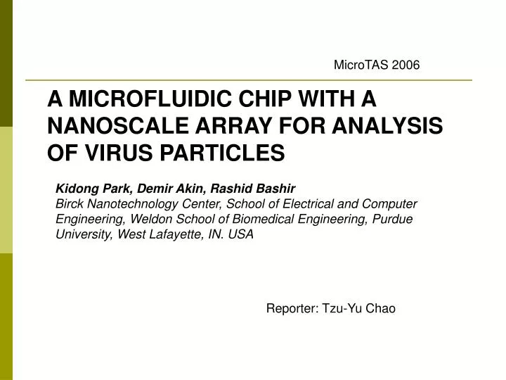
MicroTAS 2006. A MICROFLUIDIC CHIP WITH A NANOSCALE ARRAY FOR ANALYSIS OF VIRUS PARTICLES. Kidong Park, Demir Akin, Rashid Bashir Birck Nanotechnology Center, School of Electrical and Computer Engineering, Weldon School of Biomedical Engineering, Purdue University, West Lafayette, IN. USA.

E N D
MicroTAS 2006 A MICROFLUIDIC CHIP WITH A NANOSCALE ARRAY FOR ANALYSIS OF VIRUS PARTICLES Kidong Park, Demir Akin, Rashid Bashir Birck Nanotechnology Center, School of Electrical and Computer Engineering, Weldon School of Biomedical Engineering, Purdue University, West Lafayette, IN. USA Reporter: Tzu-Yu Chao
OUTLINE Introduction Method & Fabrication Experiment results Conclusion & Reference
Introduction Ref: A Scalable Addressable Positive-Dielectrophoretic Cell-Sorting Array Brian M. Taff, et al. Department of Electrical Engineering and Computer Science, Massachusetts Institute of Technology, Cambridge Ref: Manipulation of herpes simplex virus type 1 by dielectrophoresis Michael P. Hughes, et al. Bioelectronics Research Centre, Department of Electronics and Electrical Engineering, University of Glasgow, Glasgow G12 8QQ, UK
Nonmechanical filters • Sample preparation • Purification • Concentration of viral particles from a mixed sample • Real-time imaging of nanometer scale virus particles for analysis • Capture • Detection • Characterization of these particles within micro and nanoscale sensors Ref: Real-Time Virus Trapping and Fluorescent Imaging in Microfluidic Devices Demir Akin, et al. Laboratory of Integrated Biomedical Micro/Nanotechnology and Applications (LIBNA)
Method DEP force
SEM images of the probe. The gap between each probe pair is about 1-2 μm.
Experiment Result 1. polystyrene beads 2. lambda virus 3. vaccinia virus The collected 3um polystyrene beads. The beads are collected between the electrodes. (vfluid=1 mm/s, VDEP=20 Vpp @ 100 kHz)
Lambda virus captured between the probe tips. Capsid labeled by green (DiOC63, Molecular Probes) DNA labeled by red (DiL, Molecular Probes) SEM images of the captured lambda virus. (a) The probe pair with captured virus. (b) The magnified image of the lower probe.
The vaccinia viruses(牛痘病毒) were captured with positive DEP Capsid labeled by green (DiOC63) DNA labeled by blue (Hoescht33342, Molecular Probes)
Conclusion • The nano-scale probe array, integrated with micro-fluidic channel applying high electric filed, produced by its ultra sharp probe tips, the effect of the electric field on virus particle can be studied. • The capture of the virus particles is shown with colocalization of the two different fluorescence signals in real-time. References [1.] B.M. Taff & J. Voldman, A Scalable Addressable Positive-Dielectrophoretic Cell-Sorting Array, Analytical Chemistry 77, 7976-7983 (2005). [2.] D. Akin, H. Li, R. Bashir, Real-Time Virus Trapping and Fluorescent Imaging in Micro-fluidic Devices, Nano Letters, 4 (2), 257 -259, 2004. [3.] Ying Huang, et al, Differences in the AC electrodyamics of viable and non-viable yeast cells determined through combined dielectrophoresis and electrorotation studies, Phys. Med. Biol., 1992, Vol. 37, No 7, 1499~1517. [4.] M. P. Hughes, Hywel Morgan, Frazer J. Rixon, Measuring the dielectric properties of herpes simplex virus type 1 virions with dielectrophoresis, Biochimica et Biophysica Acta 1571 (2002)1-8