Download
1 / 12
120 likes | 236 Vues
Model Selection Anders Gorm Pedersen Molecular Evolution Group Center for Biological Sequence Analysis Technical University of Denmark (DTU). The maximum likelihood approach. Likelihood = Probability (Data | Model) Maximum likelihood:

E N D
Model Selection Anders Gorm Pedersen Molecular Evolution Group Center for Biological Sequence Analysis Technical University of Denmark (DTU)
The maximum likelihood approach • Likelihood = Probability (Data | Model) • Maximum likelihood: Best estimate is the set of parameter values which gives the highest possible likelihood.
Probabilistic modeling applied to phylogeny • Observed data: multiple alignment of sequences H.sapiens globin A G G G A T T C A M.musculus globinA C G G T T T - A R.rattus globin A C G G A T T - A • Probabilistic model parameters (simplest case): • Nucleotide frequencies: A, C, G, T • Tree topology and branch lengths • Nucleotide-nucleotide substitution rates (or substitution probabilities):
Computing the probability of one column in an alignment given tree topology and other parameters • A C G G A T T C A • A C G G T T T - A • A A G G A T T - A • A G G G T T T - A Columns in alignment contain homologous nucleotides Assume tree topology, branch lengths, and other parameters are given. Assume ancestral states were A and A. Start computation at any internal or external node. C A t2 t5 A A t3 t1 t4 C G Pr = C PCA(t1) PAC(t2) PAA(t3) PAG(t4) PAA(t5)
Computing the probability of an entire alignment given tree topology and other parameters • Probability must be summed over all possible combinations of ancestral nucleotides. • (Here we have two internal nodes giving 16 possible combinations) • Probability of individual columns are multiplied to give the overall probability of the alignment, i.e., the likelihood of the model. • Often the log of the probability is used (log likelihood)
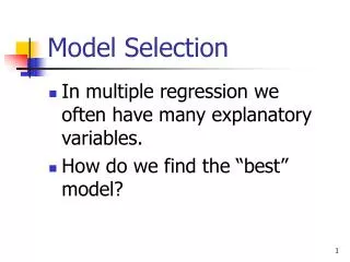
Model Selection: How Do We Choose Between Different Types of Models? Select model with best fit?
Over-fitting: More parameters always result in a better fit to the data, but not necessarily in a better description y = ax6+bx5+cx4+dx3+ex2+fx+g 7 parameter model Poor description, good fit y = ax + b 2 parameter model Good description, poor fit
Selecting the best model: the likelihood ratio test • The fit of two alternative models can be compared using the ratio of their likelihoods: LR = P(Data | M1) = L,M1 P(Data | M2) L,M2 • Note that LR > 1 if model 1 has the highest likelihood • For nested models it can be shown that = 2*ln(LR) = 2* (lnL,M1 - lnL,M2) follows a 2 distribution with degrees of freedom equal to the number of extra parameters in the most complicated model. This makes it possible to perform stringent statistical tests to determine which model (hypothesis) best describes the data
Asking biological questions in a likelihood ratio testing framework • Fit two alternative, nested models to the data. • Record optimized likelihood and number of free parameters for each fitted model. • Test if alternative (parameter-rich) model is significantly better than null-model, given number of additional parameters (nextra): • Compute = 2 x (lnLAlternative - lnLNull) • Compare to 2distribution with nextra degrees of freedom • Depending on models compared, different biological questions can be addressed (presence of molecular clock, presence of positive selection, difference in mutation rates among sites or branches, etc.)
Positive selection I: synonymous and non-synonymous mutations • 20 amino acids, 61 codons • Most amino acids encoded by more than one codon • Not all mutations lead to a change of the encoded amino acid • ”Synonymous mutations” are rarely selected against 1 non-synonymous nucleotide site CGA (Arg) CCA (Pro) CAA (Gln) ATA (Ile) CTC (Leu) 1/3 synonymous 2/3 nonsynymous nucleotide site CTA (Leu) GTA (Val) 1 synonymous nucleotide site CTG (Leu) TTA (Leu) CTT (Leu)
Positive selection II: non-synonymous and synonymous mutationrates contain information about selective pressure • dN: rate of non-synonymous mutations per non-synonymous site • dS: rate of synonymous mutations per synonymous site • Recall: Evolution is a two-step process: (1) Mutation (random) (2) Selection (non-random) • Randomly occurring mutations will lead to dN/dS=1. • Significant deviations from this most likely caused by subsequent selection. • dN/dS < 1: Higher rate of synonymous mutations: negative (purifying) selection • dN/dS > 1: Higher rate of non-synonymous mutations: positive selection
Today’s exercise: positive selection in HIV? • Fit two alternative models to HIV data: • M0: one, common dN/dS ratio in entire sequence • M3: three distinct classes with different dN/dS ratios • Use likelihood ratio test to examine if M3 is significantly better than M0, • If that is the case: is there a class of codons with dN/dS>1 (positive selection)? • If M3 significantly better than M0 AND if some codons have dN/dS>1 then you have statistical evidence for positive selection. • Most likely reason: immune escape (i.e., sites must be in epitopes) : Codons showing dN/dS > 1: likely epitopes