Download
1 / 33
420 likes | 1.14k Vues
Geometric Classification Rotator Cuff Tears. James Davidson, MD Steve Burkhart, MD Phoenix San Antonio. Goals of a Classification System. Communicate Treatment Prognosis Comparison. Previous Classifications.

E N D
Geometric ClassificationRotator Cuff Tears James Davidson, MD Steve Burkhart, MD Phoenix San Antonio
Goals of a Classification System • Communicate • Treatment • Prognosis • Comparison
Previous Classifications • Do not achieve these goals • No current Standard • Do not utilize three dimension info derived from MRI and Arthroscopy
McLaughlin Transverse Longitudinal Retracted
McLaughlin • Not widely adopted • Pre MRI • Pre Arthroscopy
DeOrio and Cofield Measure the Maximum Single Diameter Small, Medium, Large, Massive
DeOrio and Cofield • Not geometric or three dimensional
Harryman / Gerber • Number of tendons torn
Harryman / Gerber • Not geometric or three dimensional • ?? treatment • ?? prognosis
Geometric ClassificationRotator Cuff Tears A System Linking Tear Pattern to Treatment and Prognosis Arthroscopy Current Concepts In Press, 2009
Foundation • Burkhart, Adams, Arrigoni, Barth, Brady, Huberty, Lo, Parten, Pearce, Richards, Tehrani, Tauro, and others
Crescent • Short and Wide; Length ≤ Width
Crescent MRI • Length: T2 coronal • Width: T2 sagittal • L ≤ W; L < 2cm
Crescent • Repaired end to bone • Good to excellent results
Longitudinal (U’s and L’s) • Long and Narrow; Length > Width
Longitudinal MRI • Length: T2 coronal • Width: T2 sagittal • L > W; W < 2cm
Longitudinal (U’s and L’s) • Repaired side to side / margin convergence • Good to excellent results
Massive Contracted • Long and wide
Massive Contracted MRI • Length: T2 coronal • Width: T2 sagittal • L ≥ 2cm; W ≥ 2cm
Massive Contracted • Slides / Partial repair • Fair to good results
Massive Contracted • L ≥ 2cm; W ≥ 2cm most require slides/partial • L ≥ 3cm; W ≥ 3cm all require slides/partial
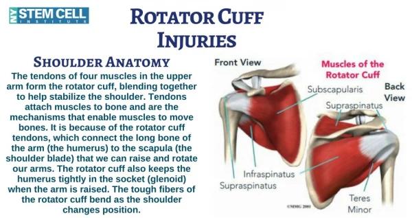
Rotator Cuff Arthropathy • Loss of Acromiohumeral Interspace • Glenohumeral Arthrosis • Irreparable by Scope or Open • Arthroplasty if Surgery
Additional NotationsRelated Pathology • Subscapularis • Biceps • Labrum • Instability • Arthritis AC or GH • Fatty Degeneration
MRI Predicts Tear Pattern Crescent Longitudinal Massive Contracted
Geometric Classification • Improved Communication • Guidance re Treatment • Guidance re Prognosis • Meaningful Comparison
Geometric Classification Thank You James Davidson, MD Steve Burkhart, MD