Download

1 / 25
250 likes | 475 Vues
Diagnosing Scoliosis C25. Objectives. What is scoliosis? What are the symptoms of scoliosis? How can scoliosis be measured? What causes scoliosis? What imaging modalities can diagnose scoliosis? What are the treatment options for scoliosis?.

E N D
Objectives • What is scoliosis? • What are the symptoms of scoliosis? • How can scoliosis be measured? • What causes scoliosis? • What imaging modalities can diagnose scoliosis? • What are the treatment options for scoliosis? https://hcavirginiaphysicians.com/about/newsroom/scoliosis-101
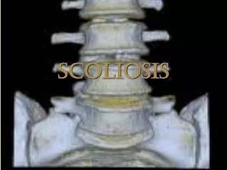
Thesis Scoliosis is a common anomaly of the spinal column, affecting two to three percent of the United States’ population.1 It can range from very minor and asymptomatic to very severe. This PowerPoint will discuss anatomy of the spine, symptoms of scoliosis, how it can be measured, causes, imaging modalities used for diagnosing and treatment options.
All About the Spine Divisions of the Spine • Divided into four regions2: • Cervical • Thoracic • Lumbar • Sacral • The thoracic and lumbar spine are the areas most commonly affected by scoliosis • Individual Vertebra • Some aspects of the individual vertebra can be altered due to scoliosis2 • Spinous process may be deviated toward one side instead of projecting straight posteriorly • Vertebral body may be oddly distorted • Lamina may be thinner due to the twisting https://slideplayer.com/slide/10499318/ https://en.wikipedia.org/wiki/Spinal_cord https://www.physio-pedia.com/Adam%27s_forward_bend_test
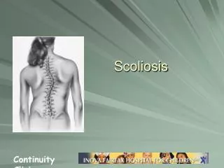
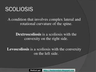
What is Scoliosis? • Most common spine malformity • Curvature of the spine • C or S curve • Any curve that measures 10° or more3 • More common in females • Develops throughout puberty4 • Types of scoliosis: • Idiopathic • Caused by a medical condition such as muscular atrophy or cerebral palsy https://www.tnchiro.com/articles/hidden-signs-of-scoliosis/
What are the Symptoms of Scoliosis? • One shoulder blade may project more posteriorly than the other4 • One hip may sit higher than the other • One arm may appear longer than the other due to the tilt of the body5 • School nurses are often the first to see signs and symptoms and will recommend seeing the doctor for measurements https://www.mymed.com/diseases-conditions/scoliosis/what-are-the-symptoms-risk-factors-and-complications-of-scoliosis https://www.treatingscoliosis.com/blog/5-most-common-scoliosis-symptoms/
How to Measure Scoliosis: Forward Bend Test • Patient will stand in front of the physician so they can observe the curve2 • Patient will bend forward • Important to keep feet together, knees straight, and hands reaching as far down as possible • A scoliometer will be placed on the shoulders to measure the severity • Determines if the scoliosis is structural or nonstructural https://www.slideshare.net/shyalachand/scoliosis-76469463 https://www.pinterest.com/pin/184577284703969917/?lp=true
How to Measure Scoliosis: Cobb Angle • Orthopedist doctor will find the most tilted vertebra at the top of the curve2 • Draw a line parallel to the vertebral end plate. • Find the most tilted vertebra at the bottom of the curve • Draw a line parallel to the inferior vertebral end plate • Draw a perpendicular line coming off of each parallel line until they connect • The angle formed is the Cobb angle • 15-20°: Observation • 20-45°: Bracing • 45-50°: Surgery https://fpnotebook.com/Ortho/T-Spine/Scls.htm
What are the Causes of Scoliosis? • Idiopathic • Unknown cause4 • Hereditary • Family history • Growth hormones • It has been found that girls with scoliosis have increased levels of growth hormones5 https://www.slideshare.net/snrifhan/idiopathic-scoliosis-72295599
Imaging Modalities for Scoliosis MRI EOS Radiography https://www.spinenevada.com/inmotion-diagnostics/scoliosis-standing-digital-xrays.html https://www.itnonline.com/content/eos-imaging-receives-fda-approval-spineeos-its-online-3d-planning-solution-spine-surgery https://pmj.bmj.com/content/86/1017/419
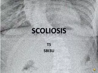
Radiography • PA or AP erect, full spine radiograph • PA is preferred to lower radiation dose to the breasts • Lateral erect • Lateral bending if deemed necessary5 • Images may be taken in the patient’s brace to determine proper fitting • Processing equipment uses a stitching method to combine upper and lower images6 • Scoliosis images are viewed in the posterior projection • Opposite of x-ray viewing
Radiography Continued • X-ray equipment uses 80-95 kVp and 40-60 mAs (depending on body habitus)7 • Young patients receive a very high dose of radiation, even with shielding • With severe scoliosis, breast shields may not be used in fear of obstructing anatomy • Images taken every 6 months8 • Women exposed to radiation during teenage years have 2-3% higher risk of developing breast cancer9 https://scoliosistreatmentalternatives.com/3668/reducing-x-ray-exposure-important-in-scoliosis-treatment-especially-with-children/
MRI • Equipment does not use radiation10 • Instead uses magnets to line up the hydrogen molecules in the body • Better for pediatrics • Exam takes about an hour10 • Harder for pediatrics to hold completely still for that long • Very loud inside the scanner • May make patients nervous and uncomfortable https://openi.nlm.nih.gov/detailedresult.php?img=PMC3116551_IJO-59-162-g003&req=4 https://www.researchgate.net/figure/MRI-T2-weighted-Coronal-Plane-Image-Idiopathic-Scoliosis-Patient-26-Schmorls-nodes-on_fig15_5629374
Advanced Technology: EOS Scanning Click here for an EOS demonstration • First scanner was installed in 2008 in Europe13 • Children’s Hospital of WI was one of the first pediatric facilities to have the new EOS scanner14 • Scanner takes two images (PA and LAT) simultaneously • Takes about 15-20 seconds to scan entire body13 • Makes 3D reformats for treatment planning • Reduces exposure by 90%8 • Used for scoliosis, growth plate fractures, leg length discrepancy, and exams that should be done weightbearing https://www.nicklauschildrens.org/medical-services/radiology/services/eos-imaging-system
EOS Scanning Continued Scanner Composition • System has two x-ray tubes that are 90° from each other and two detectors that move together11 • Total filtration of 0.1 mm Cu • Anode angle of 7° • Has a low-dose and a micro-dose setting6 Pricing • Because it is so new, it is more expensive • Purchasing, installation, and training costs = $550,000 • Digital Radiography = $167,50013 https://www.eos-imaging.us/us/patients https://www.nicklauschildrens.org/medicalservices/radiology/services/eos-imaging-system
Dose ComparisonRadiography vs. EOS • A study was done involving 132 patients with adolescent idiopathic scoliosis (AIS)6 • 99 of those patients went through imaging with the micro-dose EOS machine • The remaining 33 had imaging done with digital radiography • Thermoluminescent dosimeters (TLDs)were placed on the patients to measure entrance-skin dose at three different levels: sternal notch, nipple line, and pubic symphysis • PA images were taken and doses were compared • In conclusion, EOS performed the exam at a much lower dose rate Digital Radiography Effective dose = 67.5 μSv EOS scan Effective dose = 2.6 μSv
Pros and Cons of Radiography Pros • Does not require the purchase of new equipment • Cheaper cost • Less training for technologists • Cons • High radiation dose • Longer exam time • Uses the stitching process • Can obstruct anatomy at stitching line https://www.youtube.com/watch?v=m9HqKjGJAVQ
Pros and Cons of EOS Technology Pros • Lower dose to pediatric patients • Quicker exam time • More efficient for technologists • Cons • Very expensive • Not readily available since it is so new • Requires training for technologists https://spinalnewsinternational.com/eos-imaging-and-stryker-announce-co-promotion-agreement-for-the-uk/
Treatment Options • Observation • Bracing • Surgery • Course of treatment is determined by the likelihood of the curve progressing • The following factors lead to a higher chance of progression: • Lower Risser sign (determines skeletal maturity)14 • Stage 0 Risser sign shows no ossification of the iliac apophysis, which means the patient is still growing and developing14 • Double curves present • Larger degree of curvature • Determined by measuring the Cobb angle5 • Curves present at a young age (before menses in females) http://vsrc.com.au/what-is-a-risser-sign/
Observation • May be the only treatment necessary for small, minimal curves (15-20°)5 • Doctor may just observe if the curve has a small chance of progressing • Images taken periodically to ensure there is no progression • With age, adults who had scoliosis as a child are more likely to have chronic back pain4 https://www.shutterstock.com/search/scoliosis?studio=1
Brace • Treatment of choice for curves over 20° • Warm gauze is wrapped around the patient’s body to mold it to their exact shape and size • Many different types, shapes, and colors • Patients should follow their doctor’s orders on how many hours per day the brace should be worn • Usually only taken off for bathing • Near the end of treatment, the patient will be weaned from the brace https://www.youtube.com/watch?v=0W8egoSCY10 https://scoliosisbracinginnovations.com/for-patients/comparing-scoliosis-braces/
Surgery • Treatment option for severe curves (45-50°) • Spinal fusion • X-rays are taken before the surgery • Surgeon untwists spine and places two rods on both sides of the spine • Screws are inserted • Bone graft is placed over the spine • Grows into the spaces between the vertebrae, acting as cement15 https://www.youtube.com/watch?v=yfO7YaFH4YY http://aia5.adam.com/content.aspx?productId=117&pid=1&gid=001241
How much do you remember? What is the most common cause of scoliosis? What are the three modalities to image scoliosis? What are the three treatment options for scoliosis? Trick question! The cause of most cases of scoliosis is idiopathic (unknown). Radiography MRI EOS Scanning Observation Bracing Spinal Fusion Surgery
Conclusion • Scoliosis is a curvature of the spine with an idiopathic cause • Symptoms include uneven hips and shoulders, and one arm appearing longer than the other • The severity can be measured through the Adam Forward Bend Test and the Cobb Angle • Imaging modalities used include digital radiography, MRI, and EOS technology • EOS scanning is the newest technology being used because of its advantages • Lower dose, better for pediatrics, and a faster exam time • The treatment for scoliosis depends on the severity of the malformation • Observation, bracing, spinal fusion surgery • In the future, EOS scanners will be more widely available, lowering the radiation dose for children with scoliosis across the world
References • Scoliosis. AANS. https://www.aans.org/Patients/Neurosurgical-Conditions-and-Treatments/Scoliosis. Accessed March 21, 2019. • Scoliosis. Physiopedia. https://www.physio-pedia.com/Scoliosis. Accessed March 19, 2019. • Scoliosis of the Spine: Causes, Images, Symptoms, and Treatments. WebMD. https://www.webmd.com/back-pain/causes-scoliosis#1. Accessed March 6, 2019. • Scoliosis. Mayo Clinic. https://www.mayoclinic.org/diseases-conditions/scoliosis/symptoms-causes/syc-20350716. Published December 29, 2017. Accessed March 6, 2019. • Adolescent Scoliosis. University of Maryland Medical Center. https://www.umms.org/ummc/health-services/orthopedics/services/spine/patient-guides/adolescent-scoliosis. Accessed March 6, 2019. • Steve C. N. Hui J-PP, Judy Y. H. Wong T-ping L, Bobby K. W. Ng, Jack C. Y. Cheng, Winnie C. W. Chu. Radiation dose of digital radiography (DR) versus micro-dose x-ray (EOS) on patients with adolescent idiopathic scoliosis: 2016 SOSORT- IRSSD "John Sevastic Award" Winner in Imaging Research. Scoliosis and Spinal Disorders. https://scoliosisjournal.biomedcentral.com/articles/10.1186/s13013-016-0106-7. Published December 29, 2016. Accessed March 6, 2019. • Ho A. Scoliosis PA/AP view | Radiology Reference Article. Radiopaedia.org. https://radiopaedia.org/articles/scoliosis-paap-view. Accessed March 6, 2019. • Pediatrics. The EOS Machine for Scoliosis Monitoring. UPMC HealthBeat. https://share.upmc.com/2017/05/about-scoliosis-eos-scan/. Published August 29, 2018. Accessed March 6, 2019. • Center for Devices and Radiological Health. Resources for You (Radiation-Emitting Products) - Reducing Patient Exposure During Scoliosis Radiography. U S Food and Drug Administration Home Page. https://www.fda.gov/Radiation-EmittingProducts/ResourcesforYouRadiationEmittingProducts/ucm252765.htm. Accessed March 6, 2019. • Magnetic resonance imaging (MRI). Stanford Health Care (SHC) - Stanford Medical Center. https://stanfordhealthcare.org/medical-conditions/back-neck-and-spine/scoliosis/diagnosis/mri.html. Accessed March 8, 2019. • About us. Presentation | CONNECTING IMAGING TO CARE | EOS imaging. https://www.eos-imaging.us/us/about/history. Accessed March 8, 2019. • EOS scanner. Children. https://www.chw.org/medical-care/imaging/diagnostic-tests/x-ray/eos-scanner. Accessed March 8, 2019. • Mahboub-Ahari A, Hajebrahimi S, Yusefi M, Velayati A. EOS imaging versus current radiography: A health technology assessment study. Med J Islam Repub Iran. • Hacquebord JH, Leopold SS. In brief: The Risser classification: a classic tool for the clinician treating adolescent idiopathic scoliosis. ClinOrthopRelat Res. 2012;470(8):2335-8. • Treating scoliosis with posterior spinal fusion with instrumentation. University of Iowa Stead Family Children's Hospital. https://uichildrens.org/health-library/treating-scoliosis-posterior-spinal-fusion-instrumentation#WhatToBring. Published December 6, 2018. Accessed March 6, 2019. • Dr. Lonstein for images without citation underneath