Download
1 / 39
420 likes | 719 Vues
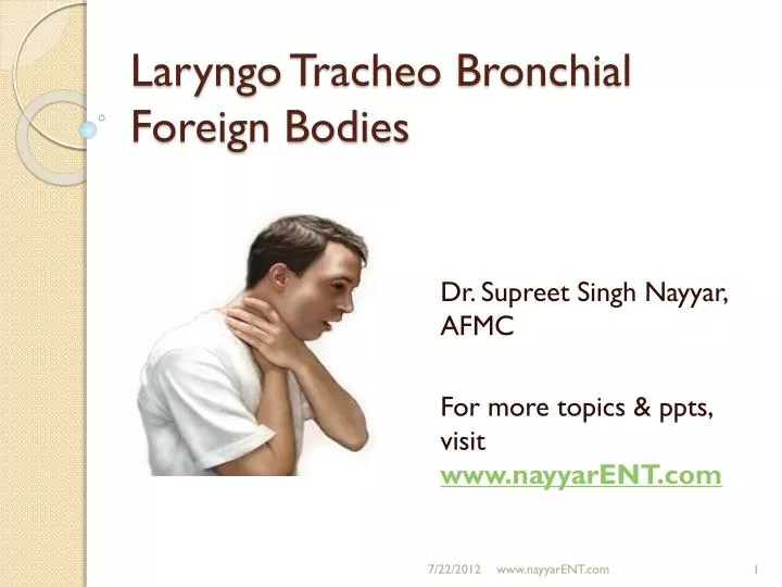
Laryngo Tracheo Bronchial Foreign Bodies. Dr. Supreet Singh Nayyar, AFMC For more topics & ppts , visit www.nayyarENT.com. Overview. Introduction Applied anatomy Aetiology Presentation Pathology Assessment Diagnosis Complications Management Post Op Care Summary References.

E N D
LaryngoTracheo Bronchial Foreign Bodies Dr. Supreet Singh Nayyar, AFMC For more topics & ppts, visit www.nayyarENT.com www.nayyarENT.com
Overview • Introduction • Applied anatomy • Aetiology • Presentation • Pathology • Assessment • Diagnosis • Complications • Management • Post Op Care • Summary • References www.nayyarENT.com
Introduction • Orifices • Curiosity of children • Minor irritation / Life threatening Problem www.nayyarENT.com
Applied anatomy Site of Lodging of Foreign Body Right Main Bronchus • The diameter of the right main bronchus is larger than the left, • The angle of divergence from the tracheal axis is smaller on the right, • Airflow through the right lung is greater than through the left, • The carina is more likely to be located to the left of midline rather than to the right. www.nayyarENT.com
Infant larynx • More anterior & higher in neck • Epiglottis larger, longer & angled more over glottis • Larynx cone-shaped: narrowest at cricoid ring • Trachea 57mm long, diam 4 mm www.nayyarENT.com
Paediatric airway • All cartilaginous supporting framework are soft, pliable & prone to collapse. www.nayyarENT.com
Rapid Subglottic Edema • Supraglottis : surrounded by loose connective tissue, prone to edema which grows rapidly • Inflammation from epiglottis can spread quickly to pre-epiglottic & para-glottic spaces. www.nayyarENT.com
Rapid Subglottic Edema www.nayyarENT.com
Aetiology • Age/Sex • Predisposing factors- • Interference with deglutition reflex • Unconscious patient • Pharyngeal / laryngeal paralysis • Improper mastication with hurried swallowing • Types • Inert / Non inert • Region www.nayyarENT.com
Presentation • Typical History immediately after aspiration • Presenting after respiratory complications www.nayyarENT.com
Pathogenesis of bronchial obstruction Stop valve Bypass valve Oneway valve Hence clinical features will vary www.nayyarENT.com
Immediate assessment • Quick history and physical examination • Vital parameters • SpO2 monitoring • ABG www.nayyarENT.com
Specific • Indirect Laryngoscopy www.nayyarENT.com
Specific • Fibreoptic Laryngoscopy www.nayyarENT.com
Specific • Direct Laryngoscopy • Fibreoptic & Rigid Bronchoscopy www.nayyarENT.com
Diagnosis • The plain chest radiography • Sensitivity 66% • Specificity 51% • Both AP & Lat view required for exact localization • May be still useful in radiolucent foreign bodies due to features of obstructive emphysema (or the ball valve mechanism) Radiology in Foreign Body www.nayyarENT.com
Radiology in Foreign Body • Radiopaque FB (23.3%)* • Hyperinflation or obstructive emphysema (21.8%)* • Hyperinflation or obstructive emphysema with atelectasis in the same hemithorax (18%)* • Lobar atelectasis (12.8%)* • Whole-lung atelectasis (6.8%)* • Shift of mediastinal shadow (11%)* • Aeration within an area of atelectasis (6%)* * Girardi G, Contador AM, Castro-Rodriguez JA.PediatrPulmonol. 2004 Sep;38(3):261-4 www.nayyarENT.com
CT Scan • Normal CT • HRCT • Reconstruction • Virtual Scopy www.nayyarENT.com
Reconstruction www.nayyarENT.com
Virtual Imaging: • Volume rendered images • Navigation beyond obstruction www.nayyarENT.com
Magnetic Resonance Imaging • Better sequences • Better characterization of lesion www.nayyarENT.com
Complications • Respiratory distress • Asphyxia • Cardiac arrest • Fever • Laryngeal edema • Pneumothorax • Hemoptysis • Pneumonia • Bronchiectasis • Bronchial stricture • Surgical emphysema www.nayyarENT.com
Emergency Management < one year: Back blows/abdominal thrusts www.nayyarENT.com
Emergency Management Small Child: Back blows www.nayyarENT.com
Emergency Management • Older Children /Adults: Heimlich manouvere www.nayyarENT.com
Emergency management • Finger Sweeping – Not recommended* • Tracheostomy might be required • * Scot Brown Otorhinolaryngology 7th Ed pg 1188 www.nayyarENT.com
Endoscopic removal Rigid bronchoscopy Fibre-optic www.nayyarENT.com
Endoscopic removal • Sniff position for aligning axes www.nayyarENT.com
Endoscopic removal • Distorted anatomy at depths • Study x-rays, lie/ diameter • Approach carefully, bleeding+ • Create forceps space • Inorg. Fbs –USUALLY TRAILING • Careful at glottis, tongue– can strip foreign body • Good bronchial toilet required www.nayyarENT.com
Endoscopic removal • Use of Fogarty catheter www.nayyarENT.com
Endoscopic removal • Flexible bronchoscopic view of a large foreign body (mini light bulb lodged in the right main bronchus of a 7-year-old boy (left, A). • The ureteral stone basket inserted through the 1.2-mm working channel of the bronchoscope has grasped the foreign body (right, B), • Proximal portion of the foreign body is pulled in to distal end of the endotracheal tube by the flexible bronchoscope (right, C). • Once the foreign body is thus secured,the entire apparatus (endotracheal tube, flexible bronchoscope, and basket with the foreign body in it) is removed en masse from the airways. www.nayyarENT.com
Endoscopic Removal • Use of laryngeal mask airway with fibreoptic bronchoscope www.nayyarENT.com
Endoscopic removal • Under fluoroscopic control • A foreign body (straight pin, arrows), aspirated into the right middle lobe of a 6-year-old girl, is seen on posteroanterior (A) and right lateral (B) radiographs • The foreign body could not be visualized by paediatric flexible bronchoscopy. It was, however, extracted by using a paediatric flexible bronchoscope and a ureteral stone forceps under fluoroscopic guidance. www.nayyarENT.com
Endoscopic Removal • New instruments – Optical Grabbing Forceps www.nayyarENT.com
Post op care • Oxygen • Watch SpO2 • Steroids • Nebulized asthalin / steroids • Chest physiotherapy www.nayyarENT.com
Summary • Most common among children • Potentially life threatening • Immediate Manouveres • Early removal to prevent oedema • Diagnosis & imaging • Endoscopes & Training • Post op care www.nayyarENT.com
References • Scott Brown ORL HNS,7th Edition • Cummings ORL HNS, 4th Edition • Gray’s Anatomy, 38th Edition • Various sources from internet (http://chestjournal.chestpubs.org) • Previous presentations on similar topics in department • Use of a Fogarty catheter for bronchoscopic removal of a foreign body.J M Wiesel, R Chisin, R Feinmesser and I Gay Chest 1982;81;524a-524 • Flexible Bronchoscopic Management of Airway Foreign Bodies in Children James P. Utz, John C. McDougall and W. Mark BrutinelChest 2002;121;1695-1700 • Retrieval of Aspirated Foreign Bodies in Children Using a Flexible Bronchoscope and a Laryngeal Mask Airway AvrahamAvital, M.D., David Gozal, M.D., KamalUwyyed, M.D.,andChaim Springer, M.D. www.nayyarENT.com
Thank youfor more topics & ppts, visit www.nayyarENT.com www.nayyarENT.com