Download
1 / 50
630 likes | 1.34k Vues
Cardiac Physiology. Cardiac Physiology - Anatomy Review. Circulatory System. Three basic components Heart Serves as pump that establishes the pressure gradient needed for blood to flow to tissues Blood vessels

E N D
Circulatory System • Three basic components • Heart • Serves as pump that establishes the pressure gradient needed for blood to flow to tissues • Blood vessels • Passageways through which blood is distributed from heart to all parts of body and back to heart • Blood • Transport medium within which materials being transported are dissolved or suspended
Functions of the Heart • Generating blood pressure • Routing blood • Heart separates pulmonary and systemic circulations • Ensuring one-way blood flow • Regulating blood supply • Changes in contraction rate and force match blood delivery to changing metabolic needs
Circulatory System • Pulmonary circulation • Closed loop of vessels carrying blood between heart and lungs • Systemic circulation • Circuit of vessels carrying blood between heart and other body systems
Cardiac Muscle Cells • Myocardial Autorhythmic Cells • Membrane potential “never rests” pacemaker potential. • Myocardial Contractile Cells • Have a different looking action potential due to calcium channels. • Cardiac cell histology • Intercalated discs allow branching of the myocardium • Gap Junctions (instead of synapses) fast Cell to cell signals • Many mitochondria • Large T tubes
Electrical Activity of Heart • Heart beats rhythmically as result of action potentials it generates by itself (autorhythmicity) • Two specialized types of cardiac muscle cells • Contractile cells • 99% of cardiac muscle cells • Do mechanical work of pumping • Normally do not initiate own action potentials • Autorhythmic cells • Do not contract • Specialized for initiating and conducting action potentials responsible for contraction of working cells
Intrinsic Cardiac Conduction System Approximately 1% of cardiac muscle cells are autorhythmic rather than contractile 70-80/min 40-60/min 20-40/min
Electrical Conduction • SA node - 75 bpm • Sets the pace of the heartbeat • AV node - 50 bpm • Delays the transmission of action potentials • Purkinje fibers - 30 bpm • Can act as pacemakers under some conditions
Intrinsic Conduction System • Autorhythmic cells: • Initiate action potentials • Have “drifting” resting potentials called pacemaker potentials • Pacemaker potential - membrane slowly depolarizes “drifts” to threshold, initiates action potential, membrane repolarizes to -60 mV. • Use calcium influx (rather than sodium) for rising phase of the action potential
Pacemaker Potential • Decreased efflux of K+, membrane permeability decreases between APs, they slowly close at negative potentials • Constant influx of Na+, no voltage-gated Na + channels • Gradual depolarization because K+ builds up and Na+ flows inward • As depolarization proceeds Ca++ channels (Ca2+ T) open influx of Ca++ further depolarizes to threshold (-40mV) • At threshold sharp depolarization due to activation of Ca2+ L channels allow large influx of Ca++ • Falling phase at about +20 mV the Ca-L channels close, voltage-gated K channels open, repolarization due to normal K+ efflux • At -60mV K+ channels close
PX = Permeability to ion X PNa 1 +20 2 PK and PCa 0 -20 PK and PCa 3 0 -40 Membrane potential (mV) PNa -60 -80 4 4 -100 0 100 200 300 Time (msec) Phase Membrane channels 0 Na+ channels open 1 Na+ channels close 2 Ca2+ channels open; fast K+ channels close 3 Ca2+ channels close; slow K+ channels open 4 Resting potential AP of Contractile Cardiac cells • Rapid depolarization • Rapid, partial early repolarization, prolonged period of slow repolarization which is plateau phase • Rapid final repolarization phase
AP of Contractile Cardiac cells • Action potentials of cardiac contractile cells exhibit prolonged positive phase (plateau) accompanied by prolonged period of contraction • Ensures adequate ejection time • Plateau primarily due to activation of slow L-type Ca2+ channels
Why A Longer AP In Cardiac Contractile Fibers? • We don’t want Summation and tetanus in our myocardium. • Because long refractory period occurs in conjunction with prolonged plateau phase, summation and tetanus of cardiac muscle is impossible • Ensures alternate periods of contraction and relaxation which are essential for pumping blood
Excitation-Contraction Coupling in Cardiac Contractile Cells • Ca2+ entry through L-type channels in T tubules triggers larger release of Ca2+ from sarcoplasmic reticulum • Ca2+ induced Ca2+ release leads to cross-bridge cycling and contraction
Electrical Signal Flow - Conduction Pathway • Cardiac impulse originates at SA node • Action potential spreads throughout right and left atria • Impulse passes from atria into ventricles through AV node (only point of electrical contact between chambers) • Action potential briefly delayed at AV node (ensures atrial contraction precedes ventricular contraction to allow complete ventricular filling) • Impulse travels rapidly down interventricular septum by means of bundle of His • Impulse rapidly disperses throughout myocardium by means of Purkinje fibers • Rest of ventricular cells activated by cell-to-cell spread of impulse through gap junctions
1 1 SA node AV node 2 1 THE CONDUCTING SYSTEM OF THE HEART SA node depolarizes. 2 Electrical activity goes rapidly to AV node via internodal pathways. SA node 3 Internodal pathways 3 Depolarization spreads more slowly across atria. Conduction slows through AV node. AV node 4 Depolarization moves rapidly through ventricular conducting system to the apex of the heart. A-V bundle 4 Bundle branches Purkinje fibers Depolarization wave spreads upward from the apex. 5 5 Purple shading in steps 2–5 represents depolarization. Electrical Conduction in Heart • Atria contract as single unit followed after brief delay by a synchronized ventricular contraction
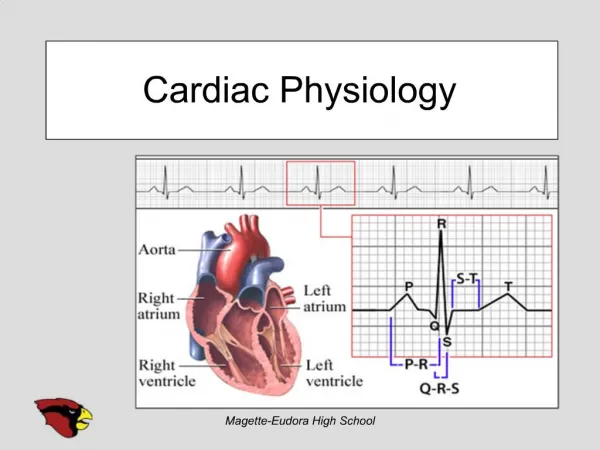
Electrocardiogram (ECG) • Record of overall spread of electrical activity through heart • Represents • Recording part of electrical activity induced in body fluids by cardiac impulse that reaches body surface • Not direct recording of actual electrical activity of heart • Recording of overall spread of activity throughout heart during depolarization and repolarization • Not a recording of a single action potential in a single cell at a single point in time • Comparisons in voltage detected by electrodes at two different points on body surface, not the actual potential • Does not record potential at all when ventricular muscle is either completely depolarized or completely repolarized
Electrocardiogram (ECG) • Different parts of ECG record can be correlated to specific cardiac events
P wave: atrial depolarization START P The end R PQ or PR segment: conduction through AV node and A-V bundle T P P QS Atria contract. T wave: ventricular Repolarization ELECTRICAL EVENTS OF THE CARDIAC CYCLE Repolarization R T P QS Q wave P Q ST segment R R wave P R Q S P R Ventricles contract. Q P S wave QS Heart Excitation Related to ECG
ECG Information Gained • (Non-invasive) • Heart Rate • Signal conduction • Heart tissue • Conditions
Cardiac Cycle - Filling of Heart Chambers • Heart is two pumps that work together, right and left half • Repetitive contraction (systole) and relaxation (diastole) of heart chambers • Blood moves through circulatory system from areas of higher to lower pressure. • Contraction of heart produces the pressure
Late diastole: both sets of chambers are relaxed and ventricles fill passively. 1 START Isovolumic ventricular relaxation: as ventricles relax, pressure in ventricles falls, blood flows back into cups of semilunar valves and snaps them closed. 5 Atrial systole: atrial contraction forces a small amount of additional blood into ventricles. 2 Isovolumic ventricular contraction: first phase of ventricular contraction pushes AV valves closed but does not create enough pressure to open semilunar valves. 3 Ventricular ejection: as ventricular pressure rises and exceeds pressure in the arteries, the semilunar valves open and blood is ejected. 4 Cardiac Cycle - Mechanical Events Figure 14-25: Mechanical events of the cardiac cycle
Wiggers Diagram Time (msec) 0 100 200 300 400 500 600 700 800 QRS complex QRS complex Electro- cardiogram (ECG) Cardiac cycle P T P 120 90 Aorta Dicrotic notch Pressure (mm Hg) Left ventricular pressure 60 Left atrial pressure 30 S2 S1 Heart sounds EDV 135 Left ventricular volume (mL) ESV 65 Atrial systole Ventricular systole Atrial systole Ventricular diastole Atrial systole Isovolumic ventricular contraction Ventricular systole Late ventricular diastole Atrial systole Early ventricular diastole Figure 14-26
KEY EDV = End-diastolic volume ESV = End-systolic volume Stroke volume 120 D ESV 80 C One cardiac cycle Left ventricular pressure (mm Hg) 40 EDV B A 0 65 100 135 Left ventricular volume (mL) Cardiac Cycle • Left ventricular pressure-volume changes during one cardiac cycle Figure 14-25
Heart Sounds • First heart sound or “lubb” • AV valves close and surrounding fluid vibrations at systole • Second heart sound or “dupp” • Results from closure of aortic and pulmonary semilunar valves at diastole, lasts longer
Cardiac Output (CO) and Reserve • CO is the amount of blood pumped by each ventricle in one minute • CO is the product of heart rate (HR) and stroke volume (SV) • HR is the number of heart beats per minute • SV is the amount of blood pumped out by a ventricle with each beat • Cardiac reserve is the difference between resting and maximal CO
Cardiac Output = Heart Rate X Stroke Volume • Around 5L : (70 beats/m 70 ml/beat = 4900 ml) • Rate: beats per minute • Volume: ml per beat • SV = EDV - ESV • Residual (about 50%)
Factors Affecting Cardiac Output • Cardiac Output = Heart Rate X Stroke Volume • Heart rate • Autonomic innervation • Hormones - Epinephrine (E), norepinephrine(NE), and thyroid hormone (T3) • Cardiac reflexes • Stroke volume • Starlings law • Venous return • Cardiac reflexes
Factors Influencing Cardiac Output • Intrinsic: results from normal functional characteristics of heart - contractility, HR, preload stretch • Extrinsic: involves neural and hormonal control – Autonomic Nervous system
Stroke Volume (SV) • Determined by extent of venous return and by sympathetic activity • Influenced by two types of controls • Intrinsic control • Extrinsic control • Both controls increase stroke volume by increasing strength of heart contraction
Stroke volume Strength of cardiac contraction End-diastolic volume Venous return Intrinsic Factors Affecting SV • Contractility – cardiac cell contractile force due to factors other than EDV • Preload – amount ventricles are stretched by contained blood - EDV • Venous return - skeletal, respiratory pumping • Afterload – back pressure exerted by blood in the large arteries leaving the heart
Frank-Starling Law • Preload, or degree of stretch, of cardiac muscle cells before they contract is the critical factor controlling stroke volume
Frank-Starling Law • Slow heartbeat and exercise increase venous return to the heart, increasing SV • Blood loss and extremely rapid heartbeat decrease SV
Extrinsic Factors Influencing SV • Contractility is the increase in contractile strength, independent of stretch and EDV • Increase in contractility comes from • Increased sympathetic stimuli • Hormones - epinephrine and thyroxine • Ca2+ and some drugs • Intra- and extracellular ion concentrations must be maintained for normal heart function
Contractility and Norepinephrine • Sympathetic stimulation releases norepinephrine and initiates a cAMP second-messenger system Figure 18.22
Modulation of Cardiac Contractions Figure 14-30
Factors that Affect Cardiac Output Figure 14-31
Medulla Oblongata Centers Affect Autonomic Innervation • Cardio-acceleratory center activates sympathetic neurons • Cardio-inhibitory center controls parasympathetic neurons • Receives input from higher centers, monitoring blood pressure and dissolved gas concentrations
Reflex Control of Heart Rate Figure 14-27
Modulation of Heart Rate by the Nervous System Figure 14-16
Establishing Normal Heart Rate • SA node establishes baseline • Modified by ANS • Sympathetic stimulation • Supplied by cardiac nerves • Epinephrine and norepinephrine released • Positive inotropic effect • Increases heart rate (chronotropic) and force of contraction (inotropic) • Parasympathetic stimulation - Dominates • Supplied by vagus nerve • Acetylcholine secreted • Negative inotropic and chronotropic effect
Regulation of Cardiac Output Figure 18.23
Congestive Heart Failure (CHF) • Congestive heart failure (CHF) is caused by: • Coronary atherosclerosis • Persistent high blood pressure • Multiple myocardial infarcts • Dilated cardiomyopathy (DCM)
Intrinsic Cardiac Conduction System Approximately 1% of cardiac muscle cells are autorhythmic rather than contractile 70-80/min Heart block 40-60/min Ectopic focus 20-40/min