Download
1 / 63
790 likes | 1.39k Vues
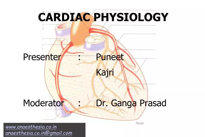
CARDIAC PHYSIOLOGY. Presenter : Puneet Kajri Moderator : Dr. Ganga Prasad. www.anaesthesia.co.in anaesthesia.co.in@gmail.com. CONTENTS. Nerve supply of the heart Neural control of heart rate Contractility – systolic and diastolic function Baroreceptor reflex Chemoreceptor reflex

E N D
CARDIAC PHYSIOLOGY Presenter : Puneet Kajri Moderator : Dr. Ganga Prasad www.anaesthesia.co.in anaesthesia.co.in@gmail.com
CONTENTS • Nerve supply of the heart • Neural control of heart rate • Contractility – systolic and diastolic function • Baroreceptor reflex • Chemoreceptor reflex • Bain bridge atrial reflex • Von Bezold-Jarisch reflex • Valsalva maneuver • Cushing reflex • Occulo cardiac reflex
NERVE SUPPLY OF THE HEART • Nerves to the heart originate from sympathetic neurons of thoracolumbar region and parasympathetic nerves originate from the cervical region.
SYMPATHETIC SYSTEM Stellate ganglion Caudal cervical sympathetic trunks Sympathetic fibers Rt. dorsal medial Dorsal lateral Large nerve Lt. main coronary artery Anterior descending Circumflex coronary
PARASYMPATHETIC SYSTEM • Parasympathetic: Preganglionic neurons arise in the medulla oblongata in the dorsal vagal nucleus ambiguous • Fibres enter the thorax as branches from the recurrent laryngeal and thoracic vagus nerves • Parasympathetic nerves are particularly abundant near the coronary sinus and SVC • Ganglia occur within the heart usually close to the structures innervated by short post ganglionic neurons
Afferent nerves from the heart ascend vial 10th cranial nerve and spinal cord to the nucleus tractus solitarius and the dorsal vagal nucleus and nucleus ambiguous to form parasympathetic motor efferent system of medullary vasomotor center • Sympathetic activation of heart originates in rostral ventrolateral medulla, ventromedial rostral medulla and parvocellular region of paraventricular nucleus
NERVE SUPPLY OF THE HEART • Both the parasympathetic and sympathetic nerves form the superficial and deep cardiac plexuses • The superficial cardiac plexus is situated below the arch of aorta in front of the right pulmonary artery, it is formed by – • The superior cervical cardiac branch of the left sympathetic chain • The inferior cervical cardiac branch of left vagus • It gives branches to • The deep cardiac plexus • The right coronary artery • The left anterior pulmonary plexus
The deep cardiac plexus is situated in front of the bifurcation of trachea and behind the arch of aorta, it is formed by - • All the cardiac branches derived from the cervical and upper thoracic ganglia of sympathetic chain • Cardiac branches of vagus and recurrent laryngeal nerves except those which form the superficial plexus • Right and left halves of plexus distribute branches to the corresponding coronary and pulmonary plexus. Separate branches are given to atria
CARDIAC RECEPTORS • Most important adrenoreceptor is heart is B1 • B2 adrenoreceptor in heart has similar cardiac effect B1 • Prejunctional a2 adrenoreceptor inhibit NE release • Prejunctional b2 adrenoreceptor facilitate NE release • Prejunctional M2 adrenoreceptor inhibit NE release • Right atrium – 74% b1 and 26% b2 • Ventricles – 86% b1 and 14% b2
NEURAL CONTROL OF HEART RATE • Dominance of sympathetic or parasympathetic system • Age • Physical condition • Inhibitory parasympathetic system usually predominates • The right stellate ganglion has effect on heart rate, the left has as more effect on contractility • Abnormalities of sympathetic cardiac nerve tone occur in long QT interval syndromes
Atrial receptors • Parasympathetic receptors type A, type B and receptors innervate by group C fibers reflexly alter intravascular volume or heart rate • Type A receptors respond to heart rate rather than atrial pressure • Type B receptors response to the atrial volume
Ventricular receptor • Baroreceptor/mechanoreceptor/sympathetic mechenosensitive or chemosensitive receptor • Postcardiotomy hypertension results from a cardiogenic reflex transmitted through the sympathetic afferent fibers of the stellate ganglion • K type, opiate receptors found in vagus, cardiac, and sympathetic ganglia, may mediate dysrhythmias, particularly during ischemia and reperfusion
SYSTOLIC FUNCTION • Traditional description of systolic function is limited to left ventricle • Systolic performance • Loading condition • Contractility • Preload and afterload – extrinsic independent factors • Preload – ventricular load at end of diastole, before contaction has started • Afterload – systolic load on the left ventricle after contraction has begun
Aortic compliance is an additional determine of after load • Preload and after load can be considered as wall stress that is present at end of diastole and LV ejection Law of laplace 6 = P X R/2H • 6 = wall stress • P = pressure • R = radius • H = wall thickness • Aortic stenosis • Heart failure
Contractility • Factors known to modify contractility are: • Max velocity of contraction (V Max) – Max velocity of ejection at zero load • V max is obtained by plotting velocity of muscle shortening in isolated papillary muscle at varying degree of force • V max cannot be measured in intact heart
To measure intrinsic contractile activity of intact heart • Best method: pressure volume loops, requiring catheterization of left side of heart • Most common: non invasive index – ejection fraction assessed by echocardiography angiography or radionucleotide ventriculography EF = (LVED – LVESV)/LVEDV
Cardiac work • External work (Stroke work): expended to eject blood under pressure • Internal work: expended within ventricle to change the shape of heart and prepare heart for ejection • Wall stress is directly proportional to internal work of heart • External work = Stroke volume x pressure SV x P (LVEDV – LVESV) x P
Clinical significance – In case of poorly drained LV external work is provided by roller pump during bypass – myocardial ischaemia still occurs because of poor drainage of LV • Efficiency of cardiac contraction is estimated by cardiac efficiency – external work/energy equivalent of O2 consumption • Heart failure • Ventricular dilatation reduce cardiac efficiency because it increase wall stress
HEART RATE AND FORCE FREQUENCY RELATIONSHIP • TREPPE or staircase phenomenon or force frequency • In isolated cardiac muscle an increase in the frequency of stimulation induces an increase in the force of contraction • Increased frequency of stimulation – incrementaly increases contraction • Lower frequency of stimulation – decreases contractile force • Extremely rapid frequency of stimulation – force of contraction decrease
Clinical significance * Pacing induced positive inotropic effects may have effective only upto a certain heart rate * In failing heart this relation may be less effective in producing a positive inotropic effect
DISATOLIC FUNCTION • Diastole is ventricular relaxation and it occurs in four distinct phases • Isovolumetric relaxation • Rapid filling phase • Slow filling or diastasis • Final filling during atrial systole • Phase • 1: does at contribute to ventricular filling • 2: maximum contribution • 3: 5% • 4: 15%
INDICES TO ASSESS DIASTOLIC FUNCTION • Index used for examining isovolumic relaxation phase is peak instantanecus rate of LV pressure decline (-dp/dt) or time constant of isovolumic LV pressure decline • Estimation of diastolic dysfunction during auxotonic relaxation – Echocardiography • Ventricular compliance can be evaluated by pressure volume relationship
Many different factors influence diastolic function • Diastolic dysfunction is decreased ability of the heart to fill • Diastolic dysfunction is now being recognized as predominant cause of CHF • In diastolic ventricular interaction dilatation of either LV or RV will have an impact on affective filling of contractive ventricle
BARO RECEPTOR REFLEX • Stretch receptors in walls of heart and blood vessels • Responsible for maintenance of blood pressure
Anatomy • Carotid-afferent nerve of Hering (glossopharyngeal) • Aortic-vagus • Cardiovascular centres in medulla Stimulus • Increased blood pressure Response • Inhibition of sympathetic and increase in parasympathetic activity, causing decreased cardiac contractility heart rate
Clinical significance • Others – Stretch receptors are activated if systemic blood pressure is greater than 170 mm of Hg • Important role during blood loss and shock, however reflex arch loses it’s functional capacity at blood pressure < 50 • Volatile anesthetics (in particular halothane) inhibit the heart rate component of this reflex • Concomitant use of CCB, ACE inhibitor or PDE inhibitor will ¯ the CVS response of BP through baroreceptor • Patients with chronic hypertension often exhibit perioperative circulatory instability as a result of ¯ in baroreceptor reflex response
CHEMORECEPTOR REFLEX • Anatomy: Carotid and aortic bodies chemoreceptors whose nerve pass through the nerve of Herring and the vagus nerve to the medullary vasomotor centres • Stimulus: Decreasing oxygen tension or increased hydrogen ion concentrations • Response: Increased pulmonary ventilation and blood pressure with decreased heart rate – stimulation of aortic bodies causes tachycardia • Other: Peripheral chemoreceptors are minimally active
CHEMORECEPTION IN CAROTID BODY CELLS • Glomus cells have Na+, Ca++, K+ channels and can fire action potentials repetitively • Voltage gated K+ channel is influenced by PO2 level K+ current is selectively inhibited by low PO2
BAIN BRIDGE ATRIAL REFLEX Anatomy • Primarily mediated through vagal myelinated afferent fibres; activation of sympathetic afferent fibers may also occur. Increased right atrial pressure directly stretches the SA node and enhances its automaticity, increasing the heart rate Stimulus • Increased vagal tone and distention of the right atrium or central veins
Response • Depends upon the preexisting heart rate • With preexisting tachycardia, there is no effect • Volume loading at a slow heart rate causes progressive tachycardia • Global atrial distention in response to high pressures causes bradycardia, hypotension, and decreased systemic vascular resistance
VON BEZOLD-JARISCH REFLEX Anatomy • Ventricular chemoreceptor and mechano-receptors with afferent pathway in unmyelinated vagal C fibres Stimulus • Noxius stimuli to either ventricle, associated with myocardial ischaemia, profound hypovolemia, coronary reperfusion, aortic steosis, neuroaxial anesthesia associated with sympathetic blockade or even vasovagal syncope
Response • Hypotension, bradycardia, parasympathetically induced coronary vasodilatation and inhibition of sympathetic outflow from vasomotor centres Clinical significance • Reperfusion of previously ischemic tissue elicits reflex • Natriuretic peptide receptors stimulated by endogenous ANP or BNP may modulate Bezold Jarisch Reflex • Thus, Bezold Jarisch reflex may be less pronounced in patients with cardiac hypertrophy or atrial fibrillation
VALSALVA MANEUVER Anatomy: • Carotid-afferent nerve of hering (glossophyrangeal) • Aortic – vagus • Cardiovascular centres in medulla Stimulus • Forced expiration against a closed glottis Response • Increased venous pressure in head, upper extremitis, with decreased right heart venous return causing decreased blood pressure and cardiac output and reflex increase in heart rate
Clinical significance • Glottic opening increases venous return to right heart, resulting in force full right and then ventricular contraction, followed by transient bradycardia • In patient with autonomic insufficiency heart rate changes are absent • Patients with primary hyperaldosteronism also fail to show the heart rate changes and blood pressure rise – reasons still obscure • Their response to valsalva maneuver returns to normal after removal of adosterone secreting tumour
CUSHING’S REFLEX • Anatomy: Increased cerebrospinal fluid pressure compresses cerebral arteries • Stimulus: Cerebral ischemia secondary to increased CSF pressure • Response: An increase in arterial pressure sufficient to reperfuse the brain • Intense sympathetic activity causes severe peripheral vasoconstriction • As a result of high vascular tone reflex bradycardia mediated by baroreceptor will ensure