Download
1 / 25
420 likes | 1.07k Vues
Reticular Formation. Dr. Sam David. Reticular Formation. Consists of neurons within the central core of the brainstem Multi-neuronal, polysynaptic pathway Receives input from almost all sensory system (except the dorsal column pathway)

E N D
Reticular Formation Dr. Sam David
Reticular Formation Consists of neurons within the central core of the brainstem Multi-neuronal, polysynaptic pathway Receives input from almost all sensory system (except the dorsal column pathway) Has efferent connections, either direct or indirect, with all levels of the CNS Hence has multiple functions and affects Motor, Sensory, Autonomic functions and responsiveness of the cortex
Reticular Formation 5 groups of Nuclei in the MEDULLA: Lateral reticular nucleus Paramedian reticular nucleus Ventral reticular nucleus Magnocellular reticular nucleus Parvicellular reticular nucleus
Lat. RN: discrete, well defined. Located near lateral surface from caudal medulla to mid olivary nucleus level. Paramedian RN: adjacent to midline. Entire length of medulla. Ventral RN: caudal 1/2 of medulla. Has small and large neurons. Caudal Medulla Oblongata
Midbrain Pons Medulla Paramedian Reticular Nucleus Ventral Reticular Nucleus Lateral Reticular Nucleus
Parvicellular RN: contains small neurons. Located immediately lateral to magnocellular nucleus. From mid-olivary level to rostral limit of medulla. Magnocellular RN: contains very large neurons Rostral Medulla Oblongata
Parvicellular Reticular Nucleus Magnocellular Reticular Nucleus Paramedian Reticular Nucleus Ventral Reticular Nucleus Lateral Reticular Nucleus
Caudal Pontine RN: extension of magnocellular nucleus Rostral Pontine RN: lacks large neurons Parvicellular RN: Located in lateral tegmentum. Extension of Parvicellular nucleus in medulla Pons
Rostral Pontine Reticular Nucleus Caudal Pontine Reticular Nucleus Parvicellular Reticular Nucleus Magnocellular Reticular Nucleus Paramedian Reticular Nucleus Ventral Reticular Nucleus Lateral Reticular Nucleus
Mesencephalic RN: Consists of scattered cells in area bounded by tectum, Red nucleus and ascending lemniscus. Midbrain
Mesencephalic Reticular Nucleus Rostral Pontine Reticular Nucleus Caudal Pontine Reticular Nucleus Parvicellular Reticular Nucleus Magnocellular Reticular Nucleus Paramedian Reticular Nucleus Ventral Reticular Nucleus Lateral Reticular Nucleus
Functional Organization Connections with the Cerebellum Red Nucleus Lateral Reticular Nucleus Spinoreticular tract Collaterals from Spinal lemniscus
Red Nucleus ICP Cerebellum Lateral Reticular Nucleus Functional Organization Connections with the Cerebellum Spinoreticular tract Collaterals from Spinal lemniscus
Functional Organization Connections with the Cerebellum Paramedian reticular nucleus Cerebellum
Connections with the Spinal Cord Motor areas of Cerebral Cortex Red Nucleus Substantia nigra Medial areas of RF Medulla & Pons Cerebellum Terminates directly or indirectly on and motor neurons SPINAL CORD
Visceral functional connections -1 Regulates visceral functions through connections with nuclei of Autonomic outflow Regulates respiration through connections with motor neurons in Phrenic nucleus and thoracic spinal cord. Reticular formation neurons involved in Respiratory & Cardiovascular control are intermingled.
Visceral functional connections -2 Maximal Inspiratory response evoked by Magnocellular Ret. Nu. in Medulla Maximal Expiratory response evoked by Parvicellular Ret. Nu. in Medulla Normal respiratory rhythm controlled by Pontine RF (Pneumotactic centre).
Visceral functional connections -3 Ventral & Magnocellular reticular nuclei have depressor effects on Heart rate and Blood pressure Lateral reticular nucleus in Medulla has opposite effects on heart rate and blood pressure.
Ascending Reticular Activating System Cortical input Olfactory input (mesencephalic RF) Medial reticular areas send collaterals that synapse with other reticular neurons. Via this repetition of relays (polysynaptic), axons reach the diencephalon ending in Hypothalamus and Thalamus (Intralaminar nuclei, and Nucleus of the midline) Superior Colliculus (retinal input) Vestibular & Cochlear Nuclei Nucleus of Spinal Tract of V Solitary Nucleus Intralaminar nuclei send fibers to other thalamic nuclei that then project to widespread areas of the cerebral cortex including non-specific association areas and areas involved in emotions. Spinal Lemniscus Spinoreticular Tract (magnocellular RF, Rostral pontine RF)
Functions of the Ascending Reticular Activating System Relatively non-specific Sensory modalities are merged in a polysynaptic pathway Only provides a vague awareness of any particular sensory modality Results in cortical stimulation with profound effects on: levels of Consciousness and Alerting reactions to sensory stimuli
Functions of the Ascending Reticular Activating System When the cortex is stimulated by the ARAS during sleep: EEG activity of the cortex changes from sleep pattern to waking state When the cortex is stimulated by the ARAS while awake: Sharpens attentiveness and creates optimal conditions for the perception of other sensory data conveyed via more direct pathways. Damage to RF results in COMA
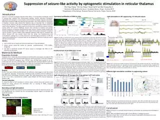
Simultaneously recorded surface EEG, EEG 1.2 mm into the cortex in a lightly anesthetized cat. Stimulation of the midbrain reticular formation (MRF) at 300 Hz at the bar produced an alerting response. (Reproduced, with permission, from Steriade M, Amzica F, Contreras D: Synchronization of fast (30–40 Hz) spontaneous cortical rhythms during brain activation. J Neurosci 1996;16:392.)
In situ hybridization images show matched coronal brain sections of the lateral hypothalamus hybridized with a 33P-labeled antisense riboprobe for orexin (a neurpeptide). Chemelli RM, et al., (1999) Narcolepsy in orexin knockout mice: molecular genetics of sleep regulation. Cell 98:437-451
(E) EEG/EMG recording during a typical narcoleptic episode. The EEG shows that the start of this episode corresponds to two high-amplitude spindling epochs, marked with arrows, associated with phasic EMG activity as muscle tone declines at the onset of attack. The star marks the onset of observable immobility. Chemelli RM, et al., (1999) Narcolepsy in orexin knockout mice: molecular genetics of sleep regulation. Cell 98:437-451
Functions of the Ascending Reticular Activating System General anesthetics are thought to suppress transmission through the RF Cutaneous stimuli and olfactory stimuli are especially important in maintaining Consciousness While visual and auditory stimuli are important in levels of alertness and attention