Download
1 / 1
10 likes | 178 Vues
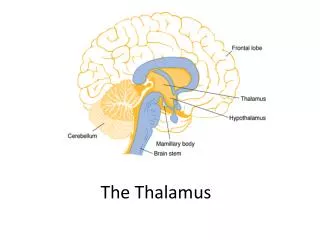
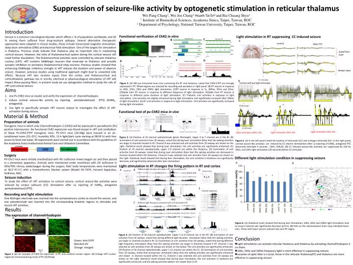
Suppression of seizure-like activity by optogenetic stimulation in reticular thalamus Wei Pang Chang 1 , Wei Jen Chang 1 Hsueh Tai En 2 and Bai Chuang Shyu 1 1 Institute of Biomedical Sciences, Academia Sinica, Taipei, Taiwan, ROC

E N D
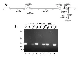
Suppression of seizure-like activity by optogenetic stimulation in reticular thalamus Wei Pang Chang1, Wei Jen Chang1Hsueh Tai En2 and Bai Chuang Shyu1 1 Institute of Biomedical Sciences, Academia Sinica, Taipei, Taiwan, ROC 2 Department of Psychology, National Taiwan University, Taipei, Taiwan, ROC F Introduction Seizure is a common neurological disorder which affects 1 % of population worldwide, and 30 % among them suffered from drug-resistant epilepsy. Several alternative therapeutic approaches were adapted in clinical studies, these include transcranial magnetic stimulation, deep brain stimulation (DBS) and electrical field stimulation. One of the targets for stimulation is thalamus. Previous study indicate that thalamus play an important role in modulating cortical seizure. However, the roles of thalamocortical system during the cortical seizure still need further elucidation. The thalamocortical activities were controlled by reticular thalamus nucleus (nRT). nRT contains GABAergic neurons that innervate to thalamus and provide synaptic inhibition on excitatory thalamocortical relay neurons. Previous studies showed that enhancing GABAergic inhibitory strength in nRT reduces the duration and power of absence seizure. However, previous studies using traditional approach might lead to unwanted side effects. Because nRT also receives inputs from the cortex, and thalamocortical and corticothalamic pathway lies in vicinity, electrical or pharmacological stimulation of nRT will impact these passing fibers. In present study we use optogenetic method to study the role of nRT and cortical seizure. Functional verification of ChR2 in vitro Light stimulation in RT suppressing CC induced seizure C Electrode in RT B Electrode in RT A A control 10Hz After PTZ 20Hz Superficial layer 50Hz 100Hz Opticfiber stimulate Recording pipette G E D Electrode in RT Electrode in thalamus 1s 50mV 50mV 1s 1s 100ms deeper layer 50ms 20ms CC stimulate B Control without light stimulate Figure 2. (A) 400 μm horizontal brain slice containing the RT and thalamus, noted that ChR2-EYFP are strongly expressed in RT. Yellow regions are selected for recording and excitation in right panel. (B) RT neuron in response to 10Hz, 20Hz, 50Hz and 100Hz light stimulation. (C)RT neuron in response to 1s, 100ms, 50ms and 20ms. (D)Spike from RT neuron in response to different frequency of light stimulation. (E)Spike from RT neuron in response to different pulse duration of light stimulation. (F) Thalamic unit activities in response to light stimulation. Unit activities are slightly enhanced during light stimulation and significantly increased after 500ms of light stimulation. (G) RT unit activities in response to light stimulation. Unit activities are significantly increased during light stimulation. ** Aims: Use Pv-ChR2 mice as model, and verify the expression of channelrhodopsin. Induce cortical seizure-like activity by injecting pentylenetetrazol (PTZ) (GABAA antagonist). Use light to specifically activate nRT neuron output to investigate the effect of nRT activation during seizure. ** ** CC stimulate Functional test of pv-ChR2 mice in vivo Material & Method Preparation of animals Transgenic mice in which only Channelrhodopsin-2 (ChR2) will be expressed in parvalbumin (Pv) positive interneurons. No functional ChR2 expression was found except in nRT and cerebellum of these Pv-ChR2-EYFP transgenic mice. PV-Chr2 mice (20-30g) were housed in an air-conditioned room (21–23°C, humidity 50%, 12-h light/dark cycle starting at 08:00 h) with free access to food and water. All experiments were carried out in accordance with the guidelines of the Academia Sinica Institutional Animal Care and Utilization Committee. thalamus response RT response CC stimulate Light stimulate 10Hz 20s Light stimulate 50Hz 20s Light stimulate 100Hz 20s Figure 3. (A) Position of 16 channel optoelectrode (green Rectangle). Upper 3 to 7 channel are in the Rt. (B) Summation of unit activities from 20 sweeps, noted that during laser stimulation (blue line) the spiking activities are larger in channels located in Rt. Channel 4 was selected and unit activities from 20 sweeps are shown on the right. Statistical result showed that during laser stimulation, the unit activities are significantly enhanced. (C) Position of 16 channel optoelectrode, upper 2-9 channel are within the thalamus. (D) Summation of unit activities from 20 sweeps, noted that during laser stimulation (blue line) the spiking activities are decreased in channel located within the thalamus. Channel 3 was selected and unit activities from 20 sweeps are shown on the right. Statistical result showed that during laser stimulation, the unit activities in thalamus are significantly decrease, and significantly enhanced after laser stimulation. Stimulation reticular thalamus(RT) Stimulation reticular thalamus(RT) Figure 5. (A) In the left panel noted the location of electrode (CC) and michigan electrode (S1). In the right panel, cortical seizure-like activates are induced by CC electric stimulatioon after i.p injecting of GABAA antagonist PTZ. (Electrode stimulate 4 seconds , 10Hz, 500uA). (B) CC induced seizure-like activates are suppressed by 100 Hz, 50Hz, and 10Hz light stimulation (20 seconds) before CC stimulate. promoter Parvalbumin Parvalbumin ChR2 Surgery PV-Chr2 mice were initially anesthetized with 4% isoflurane mixed oxygen air and then placed in a stereotaxic apparatus. Animals were maintained under anesthesia with 2% isoflurane in 30%/70% nitrous oxide/oxygen during the surgery. Rats' body temperatures were maintained at 36.5~37.5°C with a homeothermic blanket system (Model 50–7079, Harvard Apparatus, Holliston, MA). Different light stimulation condition in suppressing seizure A B Light stimulation in RT changes the firing pattern in RT and cortex Different light duration stimulate in RT and recorded in RT A B D Eletrode in RT Eletrode in Cortex di Seizure induction To study the effect of nRT activation to cortical seizure, cortical seizure-like activities were induced by corpus callosum (CC) stimulation after i.p injecting of GABAA antagonist pentylenetetrazol (PTZ). * * * E Cortical responses Recording and light stimulation One Michigan electrode was inserted into the somatosensory cortex to record the seizure, and one optoelectrode was inserted into the corresponding thalamic regions to stimulate and record nRT activities. 1000ms 250ms 750ms 500ms C Different light frequency stimulate in RT and recorded in RT Results The expression of channelrhodopsin A B cortex hippocampus Figure 6. (A) Statistical result showed that during laser stimulation, 10Hz, 20Hz and 100Hz light stimulation lead to seizure induced rate significantly decrease (p<0.5). (B) Dots on the representative brain map indicated lesion sites. Those with lower seizure induced rate near RT region. Thalamus cerebrum RT 50Hz 20Hz 100Hz 10Hz Figure 4. (A) Position of 16 channel optoelectrode. Upper 5 to 8 channel are in the RT. (B) Summation of unit activities from 20 sweeps, noted that during different light duration stimulation (blue line) the spiking activities are larger in channels located in RT. (C) Summation of unit activities from 20 sweeps, noted that during different light frequency stimulation (blue line) the spiking activities are larger in channels located in RT. Channel 2 was selected an unit activities from 20 sweeps are shown on the below. The unit activities are significantly enhanced. (D) Position of 16 channel optoelectrode, upper 1-12 channel are within the S1. (E) Summation of unit activities from 20 sweeps, noted that during laser stimulation (blue line) the spiking activities are significantly enhanced and slower in channel located within the S1. Channel 2 was selected and unit activities from 20 sweeps are shown on the right. Statistical result showed that during laser stimulation, the unit activities in thalamus are significantly enhanced, and the spiking activities pattern are slower than in RT. C D • Conclusion • Light stimulation can activate reticular thalamus and thalamus by activating channelrhodopsin-2 on them. • 10Hz, 50Hz and 100Hz frequency light is more effective in suppressing seizure. • Location of optic fiber is crucial, those in the reticular thalamus(RT) and thalamus are more effective in suppressing seizure. Green: Anti-EYFP Red:Anti-PV Orange: merged 20mm Figure 1. (A) An example of ChR2 be expressed in RT and cerebral nuclear region. (B) Enlarge nRT nuclear region.(C) Immunostaining result of PV. (D) Merge.