Download
1 / 33
430 likes | 651 Vues
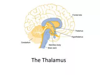
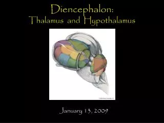
Thalamus. Dr. G. R. Leichnetz. Divisions of the Diencephalon Thalamus Hypothalamus Subthalamus : prerubral fields of Forel, zona incerta, subthalamic nucleus Epithalamus : pineal gland, habenula. EPITHALAMUS (Habenula, Pineal Gland). THALAMUS. HYPOTHALAMUS. MB.

E N D
Thalamus Dr. G. R. Leichnetz
Divisions of the Diencephalon Thalamus Hypothalamus Subthalamus: prerubral fields of Forel, zona incerta, subthalamic nucleus Epithalamus: pineal gland, habenula
EPITHALAMUS (Habenula, Pineal Gland) THALAMUS HYPOTHALAMUS MB
Coronal Section: Thalamus/ Subthalamus/ Hypothalamus Thalamus Third ventricle Subthalamus Mammillary Bodies (hypothalamus)
Thalamus/ Subthalamus/ Hypothalamus Posterior limb, internal capsule Thalamus Third ventricle Subthalamus Subthalamic nucleus Mammillary bodies (hypothalamus)
EPITHALAMUS THALAMUS Zona Incerta Subthalamic nucleus Prerubral Fields of Forel SUBTHALAMUS MB HYPOTHALAMUS
Epithalamus: Pineal Gland and Habenular Complex
Epithalamus Pineal Stalk Pineal Gland Habenular complex Pineal gland Habenular Complex Posterior commissure Pineal stalk
The epithalamus consists of the pineal gland and habenular complex. The pineal gland contains modified glial cells, called pinealocytes, that synthesize melatonin, which is released in a biological rhythm that affects activities such as sleeping and waking (jet lag). Recent evidence has shown that the suprachiasmatic nucleus of the hypothalamus (the brain’s “biological clock”), which receives direct visual input from the retina, projects to the habenular complex, which projects to the pineal gland through the pineal stalk. In the reverse direction, the pineal sends fibers to the habenular complex through the stalk to deposit melatonin in the habenula. This could represent a more direct pathway for light to influence pineal function (release of melatonin). Retina Suprachiasmatic Habenular Pineal Gland Nucleus Complex (melatonin) (“biological clock”)
The two egg-shaped thalami are joined across the third ventricle by the massa intermedia (interthalamic adhesion). The massa intermedia is not a commissure. Caudate Third ventricle Thalamus Stria terminalis Pineal gland
The two thalami are separated from each other by the third ventricle. The stria terminalis (from amygdala) runs in a groove on the floor of the lateral ventricle that separates the thalamus from the caudate nucleus. Anterior thalamic tubercle Caudate ST Thalamus IIIrd vent Pineal gland SC Pulvinar IC
The internal medullary lamina of the thalamus divides the thalamus into medial and lateral nuclear groups. The lateral nuclear group is further subdivided into dorsal and ventral tiers. DorsalTier Ventral Tier Ventral Tier
The internal medullary lamina (containing intralaminar nuclei) divides the thalamus into a medial nucleargroup(primarily MD nucleus) and lateral nucleargroup (which consists of a dorsal & ventral tiers). Medial nuclear group Lateral nuclear group Internal medullary lamina Intralaminar complex Carpenter: Human Neuroanatomy
Thalamus: Sectional Anatomy (from rostral to caudal levels)
At rostral thalamic levels: anterior nucleus is encapsulated by the internal medullary lamina; ventral anterior nucleus (VA) is in the ventral tier. Interventricular foramen of Monro Anterior thalamic tubercle ANT MTT VA MD IIIrd Vent SUBTHALAMUS
The last part of the mammillothalamic tract (MTT) from mammillary body to anterior nucleus courses through the internal medullary lamina of the thalamus. The anterior nucleus receives the MTT and projects to the cingulate gyrus. Thalamic reticular nucleus ANT Thalamic reticular nucleus MTT ANT VA MTT MD Thethalamic reticular nucleus consists of clusters of cells within the external medullary lamina of the thalamus. It is the only GABA-ergic (inhibitory) nucleus in the thalamus, and is the only thalamic nucleus that does not project to the cortex. It has intrinsic thalamic connections; involved in the generation of sleep “spindles” in the EEG.
When the MD nucleus is small, the VA nucleus is transitioning into the rostral part of the VL nucleus in the ventral tier. ANT MD VA/VL MI The massa intermedia (interthalamic adhesion) is an adhesion of ependymal and glial cells across the third ventricle (not a commissure).
When the MD nucleus is largest, the VL nucleus is present in the ventral tier. A LD/LP MD VL The mediodorsal nucleus (MD) is the largest nucleus of the medial thalamus. It receives limbic input from the amygdala and projects to the prefrontal cortex. MI The ventrolateral nucleus (VL) primarily receives input from the contralateral deep cerebellar nuclei and projects to the primary motor cortex.
In the caudal third of the thalamus, the ventral posterior nucleus (VPM/VPL) are present in the ventral tier, and the CM/Pf intralaminar nuclei are seen within the internal medullary lamina. LP PUL MD VPL CM Subthalamus Pf VPM The ventral posterolateral (VPL) and ventral posteromedial (VPM) nuclei receive the spinal and medial lemnisci. They are somatotopically organized. The centromedian (CM) and parafascicular (Pf) nuclei are the largest nucleus of the intralaminar complex. CM projects to the putamen. The Pf projects to the caudate nucleus.
The pulvinar, medial geniculate, and lateral geniculate nuclei are located in the posterior (caudalmost) thalamus. PUL Pineal gland Posterior commissure MGN Pretectum LGN The lateral geniculate nucleus receives direct visual input from the retina and projects to the primary visual cortex. The medial geniculate nucleus receives auditory input thru the brachium of the inferior colliculus and projects to the primary auditory cortex.
Basic Pattern Of Thalamic Connections All thalamic nuclei (except the thalamic reticular nucleus) have reciprocal connections with the cerebral cortex. Thalamocorticals project to lamina IV of the cortex. Corticothalamics that project back to the thalamus originate in lamina VI of the cortex. Thalamocorticals Thalamus Corticothalamics All thalamic nuclei are glutamatergic (excitatory), except the thalamic reticular nucleus (GABA). Subcortical Afferents From: Kiernan and Barr
The principal source of cortical afferents is the thalamus. Thalamocortical projections traverse the internal capsule to reach the cortex. From Noback & Demarest
The thalamus is the principal source of direct cortical afferents (corticopetal projections). The ventral tier of the lateral group contains the specific relay nuclei of the thalamus. motor relay nuclei (VA/VL) rostrally, sensory relay nuclei (VPM/VPL, MGN, LGN) caudally.
Organization of projections from the ventral tier (specific relay nuclei) to the cortex.
Corticofugal projections originate from layer V pyramidal neurons, including corticospinals, corticobulbars, corticopontines, corticostriates. Only corticothalamics originate in layer VI.There are reciprocal connections between thalamus and cortex. Cortical Efferents Associational Cortico-striate Cortico-pontine Cortico-spinal Commissural Cortico-bulbar Cortico-thalamic
The intralaminar complex is made up of subnuclei within the internal medullary lamina of the thalamus. The intralaminar nuclei receive input from the brainstem reticular formation, and project to the striatum (thalamostriates) and to the cerebral cortex (thalamocorticals). The largest of the intralaminar nuclei is the centromedian/parafascicular complex. The centromedian nucleus projects to the putamen and sensorimotor cortex. The parafascicular nucleus projects to the caudate and associational cortex.
Intralaminar nuclei are cell groups within the internal medullary lamina of the thalamus. MD MD
Intralaminar Nuclei at Caudal Thalamic Levels: CM/Pf MD Centromedian Nucleus Parafascicular Nucleus CM The centromedian and parafascicular nuclei are the largest of the intralaminar nuclei. They give rise to thalamostriates to putamen and caudate. Pf MD
The thalamic reticular nucleus is the only GABA-ergic nucleus of the thalamus. It has intrinsic thalamic connections (does not project to the cortex). Involved in generation of sleep spindles in the EEG. Thalamic reticular nucleus ANT Thalamic reticular nucleus VA MTT Intrinsic thalamic conn.’s ZI
Thalamic Pain Syndrome Central neurogenic pain (not caused by activity in peripheral sensory fibers) can be caused by lesions that interrupt the somatosensory pathway at any level. A destructive lesion that involves the ventral posterior nucleus of the thalamus may result in the thalamic pain syndrome characterized by exaggerated and exceptionally disagreeable responses to cutaneous stimulation.