Download
1 / 78
851 likes | 1.31k Vues
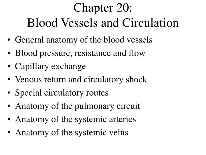
Chapter 20: Blood Vessels and Circulation. General anatomy of the blood vessels Blood pressure, resistance and flow Capillary exchange Venous return and circulatory shock Special circulatory routes Anatomy of the pulmonary circuit Anatomy of the systemic arteries

E N D
Chapter 20:Blood Vessels and Circulation • General anatomy of the blood vessels • Blood pressure, resistance and flow • Capillary exchange • Venous return and circulatory shock • Special circulatory routes • Anatomy of the pulmonary circuit • Anatomy of the systemic arteries • Anatomy of the systemic veins
Circulatory Routes • Most common route • heart arteries arteriolescapillariesvenules veins • Portal system • blood flows through two consecutive capillary networks before returning to heart • hypothalamus - anterior pituitary • found in kidneys • between intestines - liver
Circulation Routes: Anastomoses • Anastomosis = point where 2 blood vessesl merge • Arteriovenous shunt • artery flows directly into vein • fingers, toes, ears; heat loss, allows blood to bypass exposed areas during cold • Venous anastomosis • most common, blockage less serious • alternate drainage of organs • Arterial anastomosis • collateral circulation (coronary)
The Vessel Wall • Tunica externa • outermost layer • loose connective tissue • Tunica media • middle layer • usually thickest; smooth muscle, collagen, some elastic • smooth muscle for vasoconstriction and vasodilation • Tunica intima (interna) • smooth inner layer that repels blood cells & platelets • simple squamous endothelium overlying a basement membrane and layer of fibrous tissue
Arteries • Conducting (elastic) arteries - largest • pulmonary, aorta and common carotid • tunica media consists of perforated sheets of elastic tissue, alternating with thin layers of smooth muscle, collagen and elastic fibers • expand during systole, recoil during diastole; lessens fluctuations in BP • Distributing (muscular) arteries • distributes blood to specific organs; femoral and splenic • smooth muscle layers constitute 3/4 of wall thickness
Arteries and Metarterioles • Resistance (small) arteries • arterioles control amount of blood to various organs • Metarterioles • short vessels connect arterioles to capillaries • muscle cells form a precapillary sphincter about entrance to capillary
Capillaries • Thoroughfare channel - metarteriole continues through capillary bed to venule • Precapillary sphincters control which beds are well perfused • only 1/4 of the capillaries are open at a given time
Types of Capillaries • Continuous - occur in most tissues • endothelial cells have tight junctions with intercellular clefts (allow passage of solutes) • Fenestrated - kidneys, small intestine • organs that require rapid absorption or filtration; • endothelial cells have filtration pores (fenestrations) - allow passage of small molecules • Sinusoids - liver, bone marrow, spleen • irregular blood-filled spaces; some have extra large fenestrations, allow proteins and blood cells to enter
Veins • Venules • proximal venule is quite porous, exchanges fluid with tissues, like a capillary, at this point only • Venous sinuses: veins with thin walls, large lumens, no smooth muscle • Veins have lower blood pressure: avg.. 10mmHg with little fluctuation • thinner walls, less muscular and elastic tissue • expand easily, have high capacitance • venous valves aid skeletal muscles in upward blood flow
Blood Distribution, Resting Adult High Capacitance
Principles of Blood Flow • The force that the blood exerts against a vessel wall • Blood flow: amount of blood flowing through a tissue in a given time (ml/min) • Perfusion: rate of blood flow per given mass of tissue (ml/min/g) • Important for delivery of nutrients and oxygen, and removal of metabolic wastes • Hemodynamics: physical principles of blood flow based on pressure and resistance • F P/R, (F = flow, P = difference in pressure, R = resistance to flow
Blood Pressure • Measured at brachial artery of arm • Systolic pressure: BP during ventricular systole • Diastolic pressure: BP during ventricular diastole • Normal value, young adult: 120/75 mm Hg • Pulse pressure: systolic - diastolic • important measure of stress exerted on small arteries • Mean arterial pressure (MAP): • measurements taken at intervals of cardiac cycle, best estimate: diastolic pressure + (1/3 of pulse pressure) • varies with gravity: standing; 62 - head, 180 - ankle
Blood Pressure Changes With Distance More pulsatile closer to heart
Abnormalities of Blood Pressure • Hypertension • chronic resting BP > 140/90 • can weaken small arteries and cause aneurysms • Hypotension • chronic low resting BP • causes: blood loss, dehydration, anemia
Blood Pressure 2 • Importance of arterial elasticity • expansion and recoil maintains steady flow of blood throughout cardiac cycle, smoothes out pressure fluctuations and stress on small arteries • BP rises with age: arteries less distensible • BP determined by cardiac output, blood volume and peripheral resistance
Peripheral Resistance • Blood viscosity - by RBC’s and albumin • viscosity with anemia, hypoproteinemia • viscosity with polycythemia , dehydration • Vessel length • pressure and flow decline with distance • Vessel radius - very powerful influence over flow • most adjustable variable, controls resistance quickly • vasomotion: change in vessel radius • vasoconstriction, vasodilation
Laminar Flow and Vessel Radius Large radius = average velocity of flow is high Small radius = average velocity of flow is low
Peripheral Resistance • Vessel radius (cont.) • laminar flow - flows in layers, faster in center • blood flow (F) proportional to the fourth power of radius (r), F r4 • arterioles can constrict to 1/3 of fully relaxed radius • if r = 3 mm, F = (34) = 81 mm/sec; if r = 1 mm, F = 1mm/sec
Flow at Different Points • From aorta to capillaries, flow for 3 reasons • greater distance traveled, more friction to flow • smaller radii of arterioles and capillaries • farther from the heart, greater the total cross sectional area • From capillaries to vena cava, flow again • large amount of blood forced into smaller channels • never regains velocity of large arteries
Regulation of BP and Flow • Local control • Neural control • Hormonal control
Local Control of BP and Flow • Metabolic theory of autoregulation • tissue inadequatelyperfused,wastesaccumulate = vasodilation • Vasoactive chemicals • substances that stimulate vasomotion; histamine, bradykinin • Reactive hyperemia • blood supply cut off then restored • Angiogenesis - growth of new vessels • regrowthof uterine lining, around obstructions, exercise, malignant tumors • controlled by growth factors and inhibitors
Neural Control of BP and Flow • Vasomotor center of medulla oblongata: • sympathetic control stimulates most vessels to constrict, but dilates vessels in skeletal and cardiac muscle • integrates three autonomic reflexes • baroreflexes • chemoreflexes • medullary ischemic reflex
Neural Control: Baroreflex • Changes in BP detected by stretch receptors (baroreceptors), in large arteries above heart • aortic arch • aortic sinuses (behind aortic valve cusps) • carotid sinus (base of each internal carotid artery) • Autonomic negative feedback response • baroreceptors send constant signals to brainstem • BPcauses rate of signals to rise, inhibits vasomotor center, sympathetic tone, vasodilation causes BP • BPcauses rate of signals to drop, excites vasomotor center, sympathetic tone, vasoconstriction and BP
Neural Control: Chemoreflex • Chemoreceptors in aortic bodies and carotid bodies • located in aortic arch, subclavian arteries, external carotid arteries • Autonomic response to changes in blood chemistry • pH, O2, CO2 • primary role: adjust respiration • secondary role: vasomotion • hypoxemia, hypercapnia and acidosis stimulate chemoreceptors, instruct vasomotor center to cause vasoconstriction, BP, lung perfusion and gas exchange
Carotid body Aortic body Aortic body Chemoreceptors & Baroreceptors
Other Inputs to Vasomotor Center • Medullary ischemic reflex • inadequate perfusion of brainstem • cardiac and vasomotor centers send sympathetic signals to heart and blood vessels • cardiac output and causes widespread vasoconstriction • BP • Other brain centers • stress, anger, arousal can also BP
Hormonal Control of BP and Flow Angiotensin II • Angiotensinogen(prohormone produced by liver) Renin (kidney enzyme - low BP) • Angiotensin I ACE (angiotensin-converting enzyme in lungs) ACE inhibitors block this enzyme lowering BP • Angiotensin II • very potent vasoconstrictor
Hormonal Control of BP and Flow 2 • Aldosterone • promotes Na+ and water retention by the kidneys • increases blood volume and pressure • Atrial natriuretic factor ( urinary sodium excretion) • generalized vasodilation • ADH (water retention) • pathologically high concentrations, vasoconstriction • Epinephrine and norepinephrine effects • most blood vessels • binds to -adrenergic receptors, vasoconstriction • skeletal and cardiac muscle blood vessels • binds to -adrenergic receptors, vasodilation
Routing of Blood Flow • Localized vasoconstriction • pressure downstream drops, pressure upstream rises • enables routing blood to different organs as needed • Arterioles - most control over peripheral resistance • located on proximal side of capillary beds • most numerous • more muscular by diameter
Blood Flow in Response to Needs • Arterioles shift blood flow with changing priorities
Blood Flow Comparison • During exercise • perfusion of lungs, myocardium and skeletal muscles perfusion of kidneys and digestive tract
Capillary Exchange • Only occurs across capillary walls between blood and surrounding tissues • 3 routes across endothelial cells • intercellular clefts • fenestrations • through cytoplasm • Mechanisms involved • diffusion, transcytosis, filtration and reabsorption
Capillary Exchange - Diffusion • Most important mechanism • Lipid soluble substances • steroid hormones, O2 and CO2diffuse easily • Insoluble substances • glucose and electrolytes must pass through channels, fenestrations or intercellular clefts • Large particles - proteins, held back
Capillary Exchange - Transcytosis • Pinocytosis, transport vesicles across the cell, exocytosis • Important for fatty acids, albumin and some hormones (insulin)
Capillary Exchange - Filtration and Reabsorption • Opposing forces • blood (hydrostatic) pressure drives fluid out of capillary • highonarterial end of capillary, lowonvenous end • colloid osmotic pressure (COP) draws fluid into capillary (same on both ends) • results from plasma proteins (albumin)- more in blood • oncotic pressure = net COP (blood COP - tissue COP)
13 out 7 in Capillary Filtration & Reabsorption • Capillary filtration at arterial end • Capillary reabsorption at venous end • Variations • location (glomeruli- devoted to filtrationalveolar cap.- devoted to absorption • activity or trauma ( filtration)
Causes of Edema • Capillary filtration ( capillary BP or permeability) • poor venous return • congestiveheart failure - pulmonary edema • insufficient muscular activity • kidney failure (water retention, hypertension) • histamine makes capillaries more permeable • Capillary reabsorption • hypoproteinemia (oncotic pressure blood albumin) cirrhosis,famine,burns,kidneydisease • Obstructed lymphatic drainage
Consequences of Edema • Circulatory shock • excess fluid in tissue spaces causes low blood volume and low BP • Tissue necrosis • oxygen delivery and waste removal impaired • Pulmonary edema • suffocation • Cerebral edema • headaches, nausea, seizures and coma
Mechanisms of Venous Return • Pressure gradient • 7-13 mm Hg venous pressure towards heart • venules (12-18 mm Hg) to central venous pressure (~5 mm Hg) • Gravity drains blood from head and neck • Skeletal muscle pump in the limbs • Thoracic pump • inhalation - thoracic cavity expands (pressure ) abdominal pressure , forcing blood upward • central venous pressure fluctuates • 2mmHg- inhalation, 6mmHg-exhalation • blood flows faster with inhalation • Cardiac suction of expanding atrial space
Venous Return and Physical Activity • Exercise venous return in many ways • heart beats faster, harder - CO and BP • vesselsofskeletalmuscles,lungsandheartdilateflow • respiratory rate action of thoracic pump • skeletal muscle pump • Venous pooling occurs with inactivity • venous pressure not enough force blood upward • with prolonged standing, CO may be low enough to cause dizziness or syncope • prevented by tensing leg muscles, activate skeletal m. pump • jet pilots wear pressure suits
Circulatory Shock • Any state where cardiac output insufficient to meet metabolic needs • cardiogenic shock - inadequate pumping of heart (MI) • low venous return (LVR) shock - 3 principle forms • LVR shock • hypovolemic shock - most common • loss of blood volume: trauma, bleeding, burns, dehydration • obstructed venous return shock - tumor or aneurysm • next slide