Download
1 / 10
351 likes | 1.54k Vues
Carbonic Anhydrase II. Bayan K. Sheko University of Georgia Athens, Georgia December 2, 2010 . Background. Discovered in 1961 by Meldrum & Roughton Carbonic Anhydrase II is Lyase Catalyzes the reversible hydration of CO 2 H 2 O + CO 2 H + + HCO 3 -

E N D
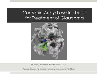
Carbonic Anhydrase II Bayan K. Sheko University of Georgia Athens, Georgia December 2, 2010
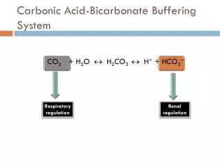
Background • Discovered in 1961 by Meldrum & Roughton • Carbonic Anhydrase II is Lyase • Catalyzes the reversible hydration of CO2 • H2O + CO2 H+ + HCO3- • The fastest enzyme 104 and 106 reactions per second.
The CA Anhydrase Family • Currently five distinct CA families exist • (α, β, γ,δ, andε) • α are mammalian CAs • β - ε CA families are highly abundant in plants, diatoms, eubacteria and archaea • no sequence homology but all contain Zinc metal • Convergent evolution
Human CA Isoenzymes • Sixteen isoenzymes (CAI-CAXVI). • Cytosolic • (CA I, CA II, CA III, CA VII, and CA XIII) • Membrane bound • (CA IV, CA IX, CA XII, and CA XIV) • Mitochondria • (CA VA, CA VB) • Secreted • CA VI • CA related proteins • (CARP), CARP VIII, CARP X and CARP XI
CA II structure Ten stranded, twisted β -sheet 30-kDa monomer Seven α helices Four minor β sheets Active site contains Zinc Figure 1. Cartoon representation of the Human CA II drawn from coordinate file 2VVA using Pymol software. Zinc metal shown in green sphere, β strands in yellow, helices in red and loops and turns in green.
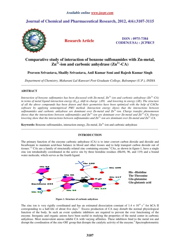
Active site Figure. 2 surface representation of CA II structure showing the relative active site. Zinc in (magenta), His 64 (yellow sticks), and His 94, 96, and 119 (red sticks). Drawn from PDB file 1TBT using pymol ( DeLano Scientific).
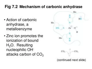
Zinc Coordination Figure 3. Schematic view of the metal center in Human CA II Showing residues that stabilize the three His residues and zinc-bound water. Figure was made from PDB code 2CBA using Pymol (Delano Scientific).
Reaction Mechanism Figure 4. Reaction mechanism of CA II. From Biochemistry. 5th edition. Berg JM, Tymoczko JL, Stryer L. New York: W H Freeman; 2002.
Histidine proton shuttle Figure 5. Histidine Proton Shuttle. From Biochemistry. 5th edition. Berg JM, Tymoczko JL, Stryer L. New York: W H Freeman; 2002.
The proton shuttle network Figure 6. The active site of HCAII depicting zinc ion tetrahedrally coordinated by His94, His96, and His119 and catalytic water 263. Hydrophobic residues are shown in the hydrophobic pocket along with deep water 338. Val 121 shown at the bottom of the active site. The proton shuttle His 64 shown in both “in” and “out’ position linked to catalytic water by water 292 and 318. Produced from PDB code 2CBA using Pymol (DeLano Scientific).