Download
1 / 65
690 likes | 738 Vues
Ashar Luqman , M.D August 10, 2010. Renal Artery Stenosis. Definition.

E N D
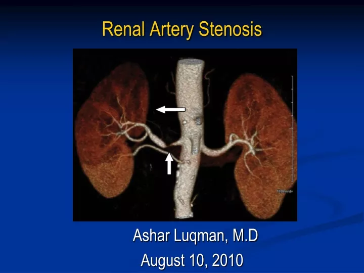
AsharLuqman, M.D August 10, 2010 Renal Artery Stenosis
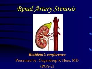
Definition Renal artery stenosis is the narrowing of the renal artery, most often caused by atherosclerosis or fibromuscular dysplasia. This narrowing of the renal artery can impede blood flow to the target kidney. Hypertension and atrophy of the affected kidney may result from renal artery stenosis, ultimately leading to renal failure if not treated Pressure gradient of 20 mmHg across stenotic area during angiography is usually considered to be of hemodynamic significance
Epidemiology Common Disease: Incidence General Population 0.1% HTN 4.0% HTN and suspected CAD 10-20% Malignant HTN 20-30% Malignant HTN and Renal Insufficiency 30-40%
Epidemiology Ultrasound screening in patients (mean age of 77): 6.8% Male : female 2:1 No racial difference (Hansen. J Vasc Surg. 2002;36:443–451) Autopsies in 221 patients > 50 years: 27% H/o diastolic HTN: 53% (Holley. Am J Med. 1964;37:14 –22) Diagnostic cath for suspected CAD: up to 20% Third most common reason of ESRD (after DM and HTN), seen in 10-20% of ESRD patients > 50 yrs old (Rihal. Mayo ClinProc. 2002;77:309 –316)
Causes Common causes Atherosclerosis Fibromuscular dysplasia Less Common causes Vasculitis (Takayasu’sarteritis) Dissection of the renal artery. Thromboembolic disease Renal artery aneurysm Renal artery coarctation Extrinsic compression Radiation injury
Fibromuscular Dysplasia Accounts for 10% of all cases of RAS. 90% cases involve media Affect females, age 15-50 Rarely leads to renal artery occlusion Genetic predisposition, smoking, hormonal factors, and disorders of the vasavasorum are possible risk factors Angiography: Site: distal renal artery Beaded, aneurysmal appearance Treatment: Ballon Angioplasty : Successful in 82-100% Restenosis in 10% Cure HTN in 60% Safian. NEJM. 2001;344: 341-442
Atherosclerosis Accounts for >2/3rd of the cases of RAS Primarily affects men over the age of 45 Usually involves the aortic orifice or the proximal main renal artery. In advanced cases, segmental and diffuse intrarenal atherosclerosis may also be observed leading to IRD This disorder is particularly common in old patients with diffuse atherosclerosis particularly with diabetes, aortoiliac occlusive disease, CAD, or HTN but can occur as a relatively isolated renal lesion 51% have progressive disease, 3-16% have total occlusion and renal atrophy developed in 21% of cases with stenosis >60% Caps MT et al Circulation 1998;98:2866
Clinical Findings HTN: Difficult-to-control hypertension despite adequate medical treatment Accelerated or malignant hypertension Severe hypertension (diastolic blood pressure >120 mm Hg) or resistant hypertension Paradoxical worsening of hypertension with diuretic therapy Onset of hypertension occurring in patients younger than 30 years or older than 50 years Hypertension with an asymmetric kidney
Clinical Findings Renal abnormalities Unexplained azotemia (suggestive of atherosclerotic renal-artery stenosis) Azotemia induced by treatment with an angiotensin-converting–enzyme inhibitor Unilateral small kidney Unexplained hypokalemia
Clinical Findings Other findings: Abdominal bruit, flank bruit, or both Severe retinopathy Carotid, coronary, or peripheral vascular disease Unexplained congestive heart failure or acute pulmonary edema
Renovascular HTN and Pulmonary Edema • Recurrent Pumonary Edema : • HTN induced increase in after-load. • Diastolic dysfunction ( LVH ) • Sodium retention, with bilateral disease. Pickering, TG, et al. Recurrent pulmonary oedema in hypertension due to bilateral renal artery stenosis: Treatment by angioplasty or surgical revascularization. Lancet 1988; 2:551.
RVH and Hyponatremia • Renovascular hypertension and hyponatremia. • Retrospective study of 32 patients with renovascular hypertension • mean plasma concentration of sodium was 129.7 meq/L, correlated inversely with plasma renin levels. • ? sec. to dipsogenic effects of angiotensin II. Agarwal, M,etal. Hyponatremic-hypertensive syndrome with renal ischemia: An underrecognized disorder. Hypertension 1999; 33:1020
Protienuria with RVH • Nephrotic range protienuria !. • Regression of protienuria with correction of vascular lesion. • Presence of protienuria, DOES NOT exlcude renovascular HTN. Chen R etal, Reversible renin mediated massive protienuria successfully treated by nephrectomy. J Urol 1995;153:133-134.
Renovascular HTN • RVH: The most common correctable cause of secondary HTN. • A syndrome of elevated blood pressure, and decreased renal perfusion. • Relatively Less common in blacks. • Incidence • ~ < 1% in patients with mild to moderate HTN. • 10-45% in patients with acute ( superimposed on essential ), severe, and or resistant HTN. Davis etal, Prevalence of renovascular hypertension in patients with grade III or IV retinopathy.N Engl J Med 1979; 301:1273
RAS and Hypertension; Possible Clinical Presentations • True reno-vascular HTN. • Essential HTN with presence of RAS, but not contributing to hypertension. • Essential HTN with superimposed HTN secondary to RAS. • RAS with presence of renal parenchymal disease. (Ischemic CKD)
Ischemic Renal Disease Critical reduction in renal blood flow to functioning renal parenchyma leading to decreased GFR, which overtime causes progressive renal injury and atrophyand thus, permanent loss of renal structural and functional integrity Ischemic renal disease has three anatomic variations: Unilateral stenosis and occlusion in a single kidney Bilateral critical renal artery stenosis or occlusion Unilateral stenosis or occluison with contralateral atrophy/nonfunction IRD Causing ESRD: 11-22%
Who should we screen? Why we need to screen?
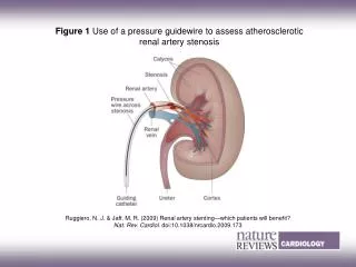
Screening And Diagnostic tests for Renal Artery Stenosis • Renal Arteriogram • Gold standard. • Non invasive Tests • Higher false negatives, especially, in patients with intra-renal involvement. • Non Invasive Tests of Choice: depends on patient factors/ availability/ expertise. • MRA • CT Angiogram • Duplex Doppler ultrasound.
MRA in Renal Artery Stenosis • Mainly for suspected atherosclerotic lesions. • Non invasive, good for proximal lesions. • Low Sensitivity for fibromuscular Disease. • Caution with GFR <30 ml/min. • Nephrogenic systemic Fibrosis: potentially fatal.
MRA and Renal Artery Stenosis Postma, CT etal Magnetic resonance angiography has a high reliability in the detection of renal artery stenosis. Am J Hypertens 1997; 10:957. • Compared MR vs Digital substraction arteriography. • N 37 • MR had 100% sensitivity, 96 % specificity . • Missed 9/12 accessory renal arteries. Vasbinder, et al. Accuracy of computed tomographic angiography and magnetic resonance angiography for diagnosing renal artery stenosis. Ann Intern Med 2004; 141:674. • 350 patients; compared MR / CTA/ DSA • 20% patients had RAS. • Sensitivity of MRA was 62% and spec. 84% • Study limitations. 36% Patients had FMD, large no. of patients without high clinical suspicion. • MR has limited utility in FMD (22 % sensitivity. 96% specificity.)
Spiral CT with CT Angiogram: • Non-Invasive, highly accurate, lower risk than Digital Substraction Arteriography . • Nephrotoxic potential. • Better for atherosclerotic lesions, lower sensitivity In FMD ~ 28%. • Kim, et al. Renal artery evaluation: comparison of spiral CT angiography to intra-arterial DSA. J Vasc Interv Radiol 1998; 9:553. • N 50 • Senstivitiy of 90 and spec. 97% for lesions greater than 50%. • Senstivity 100 and specificity 97 (patients with main renal artery disease ) • Olbricht, et al. Minimally invasive diagnosis of renal artery stenosis by spiral computed tomography angiography. Kidney Int 1995; 48:1332 • N 62, spiral CT with Angiogram compared with arteriogram. • 98% sensitive. 94 %specific.
Duplex Doppler Ultrasound • Non Invasive, can detect bilateral disease/ recurrent and or re-stenosis. • Anatomic and functional assessment of renal arteries. Compares systolic flow velocity of renal artery to aorta./ increase in end diastolic velocity in post stenotic area. • Highly operator dependant/ time consuming . • Measurement of peak systolic velocity 85% sensitive and 92% specific. Williams, GJ, et al. Comparative accuracy of renal duplex sonographic parameters in the diagnosis of renal artery stenosis: paired and unpaired analysis. AJR Am J Roentgenol 2007; 188:798.
Tests no more Recommended • Plasma rennin avtivity, captoprilrenogram , and intravenous pyelogram • Not currently recommended for screening, b/c of low senstivity, high false negative rates. • Oral captopril 25-50 mg is given, prior to isotope injection Major criteria for positive test. • Decreased relative uptake with one kidney accounting for <40% of total GFR. • Delayed peak uptake of the isotope 10-11 min. • Slower washout in stenotic kidney. ( > 5 min. delay)
CLINICAL SIGNIFICANCE OF STENOTIC LESIONS. Clinical history is important. >75% Occlusion of 1 or both arteries on arteriogram is diagnostic. 50% stenosis with post-stenotic dilatation is suggestive of RVH.
Predicting outcomes of the revascularization • Resistive Index: • 1-end-diastolic velocity/peak systolic velocity • 6000 Patients with HTN/ and clinical suspicion, screened for RVH., • 131 had RAS, . • Patients with resistive index >0.8 • 80% had decline in renal function • 50% dialysis dependant • Only 1 patient had > 10mmhg reduction in BP. • Patients with RI < 0.8 • 94 % had significant reduction in BP. • 3% had decline in renal function Radermacher, J, et al. Use of Doppler ultrasonography to predict the outcome of therapy for renal-artery stenosis. N Engl J Med 2001; 344:410.
Predicting outcomes of the revascularization Radermacher, J, et al. Use of Doppler ultrasonography to predict the outcome of therapy for renal-artery stenosis. N Engl J Med 2001; 344:410.
Predicting outcomes of the revascularization Radermacher, J, et al. Use of Doppler ultrasonography to predict the outcome of therapy for renal-artery stenosis. N Engl J Med 2001; 344:410.
Treatment Recommendations are based on 2005 ACC/AHA guidelines for peripheral arterial disease.
Medical Therapy • Optimal medical therapy in recent randomized trials has included aggressive blood pressure (BP) control with multiple agents, lipid lowering therapy with a statin, antiplatelet agents and smoking cessation • ACE-I, CCB, BB (Level of evidence A) • ARB ( Level of Evidence B) • Concern with ACE-I • Reduction In GFR. • Usually minimal elevation in SCr., only 5-10% have larger increase • Severe bilateral disease: Marked reduction in GFR with ANY HTN agent. -- intervention recommended
PTRA and Stenting for RAS • 2005 ACC/AHA Guidelines: • StentingOstial lesions.(patients meeting criteria for intervention) • Balloon angioplasty alone for FMD ( with bailout stent , if necessary)
PTRA and Stenting for RAS • Angioplasty alone is less successful. • Meta-analysis of 24 studies comapring Angioplasty vs stent placenment at 6-29 months Restenosis: 26% VS 17% Tech. success: 77% vs 98% • Small non randomized studies shown benefit for • BP control and improvement in renal function • However those studies were not well powered HighlyEffective in Ostial lesions. • 65-80% success rate. 11-17% re-stenosis.
Surgical Revascularization • Surgical Revascularization : • Bilateral Repair, unilateral repair with contralateralnephrectomy of non functioning , atrophic kidney. • 70-90% cure rate reported. • 3-6% mortality. • Indications: Aortic Reconstruction ( Aneurysm, aorto-iliac occlusive disease ) Multiple small renal arteries. Stanley JC, Surgical Treatment of renovascular hypertension. Am J Surg 1997;174:102-110)
Surgical Revascularization Vs Other Therapies • 42 prospective or retrospective cohort studies of either endovascular or surgical treatment of ARAS documented 30-day mortality rates that were 3.1% higher(1.8%–4.4%) among surgically treated patients, despite the fact that these patients who underwent surgical repair were younger (62 versus 68 years), less likely to have IHD (53% versus 60%), and less likely to have DM (14% versus 26%) Abela R, et. An analysis comparing open surgical and endovascular treatment of atherosclerotic renal artery stenosis. Eur J VascEndovasc Surg. 2009;38:666-675 • Uzzo et al compared medical therapy with surgery: • 52 pts, with ckd, uncontrolled HTN and >75% RAS in any one of renal artery • No difference in prim outcome (HTN, worsening Cr, CV endpoint) after 6 yr f/u • Balzer et al compared 50 ARAS complications with angioplasty vs surgery • Surgery was associated with non significant (25% vs 18%) in 4 yr f/u
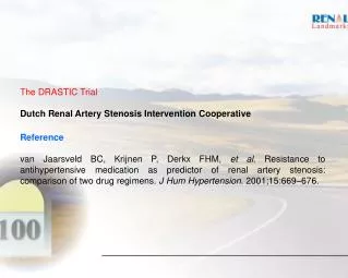
Revascularization VS Medical Therapy Randomized controlled trials DRASTIC ( Dutch renal artery stenosis Intervention co-op trial) Apr 2000 NEJM STAR ( Stent placement Atherosclerotic renal artery trial) : June 2009 Ann Int Med ASTRAL ( Angioplasty and stent in Renal artery Lesions): Nov 2009 NEJM CORAL ( CV outcomes in Renal Atherosclerotic Lesions): Enrollment finished in Jan 2010, follow up will cont till 2014
DRASTIC Prospective, randomized study was conducted at 26 centers in the Netherlands between January 1993 and November 1998 Renal Arteriography done in 543 patients because their diastolic blood pressure, measured at three consecutive visits was at least 95 mm Hg despite treatment with a standardized regimen of two antihypertensive drugs or because their serum Cr on the second or third visit had risen by at least 0.2 mg/dl during treatment with an ACE
DRASTIC 169 were found to have ostial or nonostial renal-artery stenosis (defined as a decrease in luminal diameter of more than50 percent) and thus were considered candidates for the treatment phase Out of those 63 were excluded because of Single functioning kidney and a serum Cr greater than 1.7 mg per deciliter Affected kidney that was less than 8.0 cm long, as determined by USG Total occlusion of the renal artery Aortic aneurysm necessitating surgery; Renal-artery stenosis due to fibromuscular dysplasia 106 were eligible for the treatment phase and were randomly assigned to undergo balloon angioplasty or to receive antihypertensive-drug therapy