Download
1 / 29
360 likes | 859 Vues
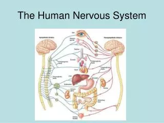
The Human Nervous System. Figure 22.1. The Nervous System. Meninges protect brain and spinal cord Dura mater: Outermost layer Arachnoid mater: Middle layer Subarachnoid space contains cerebrospinal fluid (CSF) Pia mater: Innermost layer Blood–brain barrier. The Meninges and CSF.

E N D
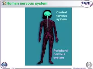
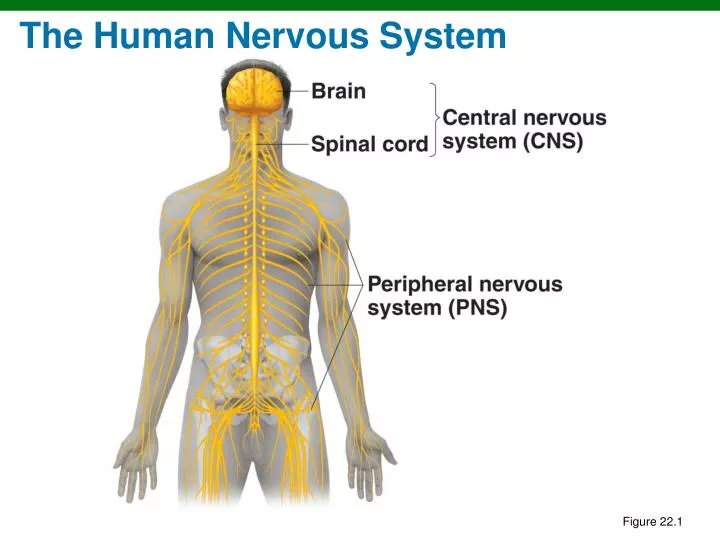
The Human Nervous System Figure 22.1
The Nervous System • Meninges protect brain and spinal cord • Dura mater: Outermost layer • Arachnoid mater: Middle layer • Subarachnoid space contains cerebrospinal fluid (CSF) • Pia mater: Innermost layer • Blood–brain barrier
The Meninges and CSF Figure 22.2
The Nervous System • Meningitis: Inflammation of meninges • Encephalitis: Inflammation of the brain • Meningoencephalitis: Inflammation of both
Bacterial Meningitis • Initial symptoms of fever, headache, and stiff neck • Followed by nausea and vomiting • May progress to convulsions and coma • Diagnosis by Gram stain and latex agglutination of CSF • Treatment: Cephalosporins, vancomycin
Spinal Tap (Lumbar Puncture) Figure 22.4
Haemophilus influenzae Meningitis • Occurs mostly in children (6 months to 4 years) • Gram-negative aerobic bacteria, normal throat microbiota • Capsule antigen type b • Prevented by Hib vaccine
Neisseria Meningitis • Also called meningococcal meningitis • Caused by N. meningitidis • Gram-negative, aerobic cocci with a capsule • 10% of people are healthy nasopharyngeal carriers • Begins as throat infection, rash • Serotypes B, C, Y, W-135 in U.S. • Serotype B in Europe • Serotype A in Africa, China, and Middle East • Vaccination (B, C, Y, W-135 capsule) recommended for college students
Neisseria Meningitis Figure 22.3
Streptococcus pneumoniae Meningitis • Also called pneumococcal meningitis • Caused by S. pneumoniae (a gram-positive diplococcus) • 70% of people are healthy nasopharyngeal carriers • Most common in children (1 month to 4 years) • Mortality: 30% in children, 80% in elderly • Prevented by vaccination
Listeriosis • Caused by Listeria monocytogenes • Gram-negative aerobic rod • Usually foodborne; it can be transmitted to fetus • 1600 cases annually in the US Figure 22.5
Tetanus • Caused by Clostridium tetani • Gram-positive, endospore-forming, obligate anaerobe • Grows in deep wounds • Tetanospasmin released from dead cells blocks relaxation pathway in muscles • Prevention by vaccination with tetanus toxoid (DTP) and booster (dT) • Treatment with tetanus immune globulin
An Advanced Case of Tetanus Figure 22.6
Botulism • Caused by Clostridium botulinum • Gram-positive, endospore-forming, obligate anaerobe • Intoxication comes from ingesting botulinal toxin • Botulinal toxin blocks release of neurotransmitter, causing flaccid paralysis • Prevention • Proper canning • Nitrites prevent endospore germination in sausages
Botulism • Treatment: Supportive care and antitoxin • Infant botulism results from C. botulinum growing in intestines • Wound botulism results from growth of C. botulinum in wounds
Botulinal Types • Type A toxin • 60–70% fatality • Found in CA, WA, CO, OR, NM • Type B toxin • 25% fatality • Europe and eastern United States • Type E toxin • 25% fatality • Found in marine and lake sediments • Pacific Northwest, Alaska, Great Lakes area
Leprosy • Also called Hansen’s disease • Caused by Mycobacterium leprae • Acid-fast rod that grows best at 30°C. • Grows in peripheral nerves and skin cells • Transmission requires prolonged contact with an infected person
Leprosy • Tuberculoid (neural) form: Loss of sensation in skin areas; positive lepromin test • Lepromatous (progressive) form: Disfiguring nodules over body; negative lepromin test
Leprosy Lesions Figure 22.9a
Leprosy Lesions Figure 22.9b
Poliomyelitis (Polio) • Poliovirus • Transmitted by ingestion • Initial symptoms: Sore throat and nausea • Viremia may occur; if persistent, virus can enter the CNS • Destruction of motor cells and paralysis occurs in <1% of cases • Prevention: vaccination (enhanced-inactivated polio vaccine)
Worldwide Annual Incidence of Poliomyelitis Figure 22.11
Rabies • Caused by the rabies virus • Transmitted by animal bite • Furious rabies: Animals are restless then highly excitable • Paralytic rabies: Animals seem unaware of surroundings Clinical Focus, p. 625
Pathology of Rabies Infection Figure 22.12
Reported Cases of Rabies in Animals Figure 22.13
Reported Cases of Rabies in Animals Figure 22.13
Rabies Virus • Virus multiplies in skeletal muscles and then brain cells, causing encephalitis • Initial symptoms may include muscle spasms of the mouth and pharynx and hydrophobia
Cryptococcus neoformans Meningitis • Also called cryptococcosis • Soil fungus associated with pigeon and chicken droppings • Transmitted by the respiratory route; spreads through blood to the CNS • Mortality up to 30% • Treatment: Amphotericin B and flucytosine
Cryptococcus neoformans Figure 22.15