Download
1 / 86
860 likes | 871 Vues
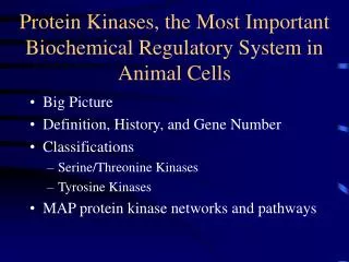
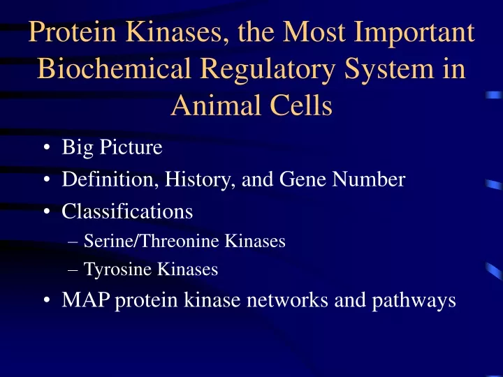
Protein Kinases, the Most Important Biochemical Regulatory System in Animal Cells. Big Picture Definition, History, and Gene Number Classifications Serine/Threonine Kinases Tyrosine Kinases MAP protein kinase networks and pathways. Sutherland Second Messenger Hypothesis.

E N D
Protein Kinases, the Most Important Biochemical Regulatory System in Animal Cells • Big Picture • Definition, History, and Gene Number • Classifications • Serine/Threonine Kinases • Tyrosine Kinases • MAP protein kinase networks and pathways
Protein Kinases in Animal Cells • Cell division • Apoptosis The first messenger interacts with a receptor and a second messenger is formed
History and Importance I • About 518 genes in humans encode protein kinases; there are an estimated 30,000 genes in humans, so that about 1.7% of the human genome encodes protein kinases • Protein kinases are the fourth largest gene family in humans • C2H2 zinc finger proteins (3%) • G-protein coupled receptors (2.8%) • Major histocompatibility (MHC) complex protein family (2.8%)
How Many Protein Kinases Are There? (518) • The kinome refers to all protein kinases in the genome • There are 478 conventional eukaryotic protein kinases (ePKs) plus 106 pseudogenes • 388 Protein-serine/threonine kinases • 90 Protein-tyrosine kinases • 58 receptor PTKs • 32 Non-receptor PTKs • There are 40 atypical protein kinases (e.g. EF2K/alpha kinases) • 478 + 40 = 518 • The exact numbers aren’t important; understand the classification
General Classes I • ACG Group • Protein kinase A; cyclic AMP-dependent protein kinase • Protein kinase C • Protein kinase G • Basic amino acid-directed enzymes that phosphorylate serine/threonine (you don’t have to memorize any sequences)
General Classes II • CaMK • Calcium-calmodulin-dependent protein kinases • I • II • III • IV • Type II is a broad specificity kinase • The others are dedicated kinases with a limited substrate specificity
General Classes III • CMGC • Cyclin-dependent protein kinases • These are important regulators of the cell cycle • MAP (Mitogen activated protein/microtubule associated protein) kinases • Many of these promote cell division • GSK3 (glycogen synthase kinase-3) • Clk (Cyclin-dependent like kinase)
General Classes IV • PTK (Protein-tyrosine kinases) • Receptor, e.g., epidermal growth factor receptor, insulin receptor • Non-receptor, e.g., Src, Abl protein kinases • Specifically phosphorylate protein-tyrosine (note they are not tyrosine kinases but protein-tyrosine kinases)
Protein Kinase Classifications • Protein-Serine/threonine • Protein-Tyrosine • Receptor: ligand binding domain and catalytic site on the same polypeptide • Non-receptor: catalytic domain separate from the receptor • Dual Specificity (both serine/threonine and tyrosine) also occur • Broad Specificity: have several substrates, e.g., PKA • Narrow Specificity: have one or a few substrates, e.g., pyruvate dehydrogenase kinase with one substrate • Classified by activator: PKA, PKG, PKC
Reactions and Types of Protein Kinases • Be able to recognize the three amino acids with an –OH in their R-group • Serine • Threonine • Tyrosine
Activation of Protein Kinases • Know PKA activation mechanism • It is the only one where there is a dissociation of regulatory subunits from catalytic subunits • This was the first activation mechanism to be described, but it turns out to be atypical or unique • PKG, allosteric • PKC, allosteric
Hormones and cAMP Many first messengers lead to changes in [cAMP]
Protein Kinase A Structure • Bilobed • N-lobe (upper) mostly beta sheet • C-lobe (lower) mostly alpha helix • Active site between the two lobes • ATP is bound in the active site
PKA Substrate Specificity Basic-Basic-Xxx-Ser-Hydrophobic is preferred
Selected Protein Kinase A Substrates • Phosphorylase kinase alpha and beta subunits • Pyruvate kinase (Liver type) • 6-Phosphofructo-2-kinase/phosphatase • Hormone sensitive lipase • Protein phosphatase inhibitor 1 • CREB (a transcription factor) • Aromatic amino acid hydroxylases • Tyrosine hydroxylase • Tryptophan hydroxylase • Phenylalanine hydroxylase • What else is special about these three enzymes? • Raf • Grandmother
Earl W. Sutherland, Jr (cAMP)Edwin Krebs (PKA) Albert G. Gilman (G-protein)Edmund Fischer (PKA) Martin Rodbell G-protein)
Enzyme Cascades and Phosphorylase and Synthase • Hormonal regulation • Hormones (glucagon, epinephrine) activate adenylyl cyclase • Glucagon, liver • Epinephrine in muscle • cAMP activates kinases and phosphatases that control the phosphorylation of phosphorylase and glycogen synthase • GTP-binding proteins (G proteins) mediate the communication between hormone receptor and adenylyl cyclase • Learn the regulation of the PKA-phosphorylase cascade!!!
Regulation of Glycogen Metabolism PKA phosphorylase kinase phosphorylase A kinase acting on a kinase that phosphorylates a protein is a cascade Steps 3 and 4 make up the first cascade to be described
Protein Kinase G • Activated by cGMP • Second second messenger protein kinase (after cAMP) • Little known about physiological protein substrates despite extensive investigation • Two types of guanylyl cyclase • Second messenger generated by atrial naturetic factor as an integral membrane guanylyl cyclase • Second messenger generated by NO action on the soluble guanylyl cyclase reaction
Action of cGMP Review Fig 19-23 for NO biosynthesis
Cyclic GMP Metabolism (Fig. 19-24) • Integral membrane guanylyl cyclase (ANF {atrionaturetic factor} receptor) • Soluble: Activated by NO
Selected Protein Kinase G Substrates • G substrate (cerebellum) • Vasodilator-stimulated phosphoprotein • Most substrates and their functions are unknown
Calcium-Dependent Protein Kinases • Protein Kinase C • Requires calcium, diacylglycerol, and phospholipid for activity • Diacylglycerol generated by the action of phospholipase C • Activated by phorbol esters (tumor promoters) • Many isozymes • Calcium-calmodulin-Dependent Protein Kinases • CAM Kinases I, II, III, IV • Several other protein kinases activated by calmodulin including myosin-light chain kinase, phosphorylase kinase, some isoforms of adenylyl cyclase and some isoforms of phosphodiesterase
Hormones and Phospholipase C • Heterotrimeric Gq activates PLC No need to memorize any of these
Hydrolysis of PIP2 • Two second messengers are generated • DAG activates PKC • IP3 leads to a rise in cytosolic Ca2+ • This activates PKC and CaM Kinases • Fig. 12-6
Protein Kinase C Family • PKC • C refers to calcium • DAG and phospholipid were also described as necessary for the activation of this enzyme • There are many isozymes that are products of different genes • It is paradoxical to have PKCs that are independent of Ca2+ and DAG
Selected Protein Kinase C Substrates • Glycogen synthase (at least two sites, inactivates) • PDGF receptor • EGF receptor • Insulin receptor • Transferrin receptor • Ribosomal protein S6 • Raf • No need to memorize any of these
Calcium-calmodulin Dependent Protein Kinases • CAM Kinase I: synapsin I and II • CAM Kinase II • CAM Kinase II (autophosphorylation) • Tyrosine hydroxylase (rate-limiting for catecholamine biosynthesis) • CREB transcription factor • Many others • CAM Kinase III: Elongation factor II of protein synthesis (This is a dedicated protein kinase) • CAM Kinase IV • Found in the nucleus • Also phosphorylates the CREB transcription factor • Many others
Receptor Protein-Ser/Thr LigandsTransforming Growth Factor-β Ligands • This family has diverse functions (that you don’t need to remember) • BMP2 subfamily: BMP2 and BMP4, chondrogenesis and other developmental functions • BMP5 subfamily: BMP5,6,7,and 8, development of nearly all organs • BMP3/osteogenin subfamily: bone formation • Activin subfamily: erythroid cell differentiation • TGF-β1, 2, and 3: control of proliferation and differentiation; production of the extracellular matrix • There are about 15 BMPs • They are synthesized as integral membrane proteins and the BMP is cleaved extracellularly in a regulated fashion • Proteins contain about 450 aa; BMPs are about 110 residues • BMP-2 and BMP-4 are expressed by human adult pulp tissue • BMP 1 is a C-terminal procollagen protease
Smads • Transforming growth factor beta ligands including the BMPs activate receptor protein Ser/Thr kinases • Smad transcription factors are phosphorylated and activated • Smads enter the nucleus to bring about a response • These human proteins are homologous to Drosophila proteins called Sma or Mad, thus smad • Smads play a role in normal cell growth, cell division, and apoptosis (programmed cell death)
Protein-Tyrosine Kinases • Receptor • Insulin • Epidermal Growth Factor • Platelet Derived Growth Factor • Non-receptor • Src protein kinase • Abl and Bcr-Abl • Jak (Janus kinase [two catalytic regions]) or whimsically, just another kinase
Human Protein-Tyrosine Kinases from the Human Genome Project • The protein-tyrosine kinases are a large multigene family with particular relevance to many human diseases, including cance • A search of the human genome for tyrosine kinase coding elements identified several novel genes and enabled the creation of a nonredundant catalog of tyrosine kinase genes • Ninety unique kinase genes can be identified in the human genome, along with five pseudogenes • Of the 90 tyrosine kinases, 58 are receptor type, distributed into 20 subfamilies • The 32 nonreceptor tyrosine kinases can be placed in 10 subfamilies
Epidermal Growth Factor Receptor Family • In the 1950s-60s Stanley Cohen discovered a factor present in crude submaxillary preparations that induced precocious tooth development in newborn mice that he called epidermal growth factor • He purified EGF, determined its sequence, studied its binding to the EGF receptor, showed that a single molecule contained EGF binding and protein kinase activity • He demonstrated that the EGF receptor was a protein-tyrosine kinase, the first to be described (1980) • He also showed that EGF and receptor are taken up by cells and are degraded in lysosomes (many receptors undergo this fate)
EGF Growth Factor and Receptor Family • Null mutants of any family member are embryonic lethal • Important in development • Implicated in many cancers
ErbB and Malignancies • Head and neck squamous cell carcinomas (>90% associated with ErbB overexpression) • 27,000 new cases in the US per year • Bladder • Breast • Kidney • Non-small cell lung • Prostate cancers • Many other solid tumors
CA of the Tongue • Early squamous cell carcinoma of the tongue • Malignant neoplasms of the oral cavity account for 3-5% of all malignancies • 50% involve the tongue (lateral border and ventral surface most common) • Then floor of the mouth > gingiva > alveolar mucosa > buccal mucosa > palate • Squamous cell carcinomas account for >90% of all malignancies of the oral cavity • Men/woman = 2/1 • Usually more than 40 years of age • Under diagnosed; more than 50% have metastasized at the time of Dx
Receptor Activation • Ligand binds, dimers form, transphosphorylation occurs, and the receptor is activated • Homodimers: ErbB1/ErbB1, etc. • Heterodimers: ERbB1/ErbB2 (common in breast cancer) • One of the dimers phosphorylates the other, and the other dimer phosphorylates the one
Monoclonal Abs in the Rx of Cancer • Mabs are directed toward the ectodomain of the ErbB2/HER2 receptor • Herceptin • 20-30% of all human breast cancers overexpress ErbB2, or HER2 (Human Epidermal growth factor Receptor) • These tumors can be treated with Herceptin • It targets domain IV of ErbB2 • Erbitux • Treatment of colorectal cancer that has spread • In combination with irenotecan (a DNA topoisomerase I inhibitor) • From ImClone (The Martha Stewart case) • Approved by US FDA in February 2004
Structure of the EGF Receptor Protein-Tyrosine Kinase Domain • Open activation loop is active (blue or green) • Compact activation loop is inactive (magenta) • This is an important regulatory concept • Blue: EGF unphosphorylated • Green: IRK phosphorylated • Magenta: IRK unphosphorylated