Download
1 / 1
10 likes | 107 Vues
Figure 4. Changes in the localization of Mitochondria with age in EDL muscle (representative images). Figure 1. Changes in the expression of 4-HNE in with age in Soleus muscle (representative images). Figure 2. Changes in the expression of

E N D
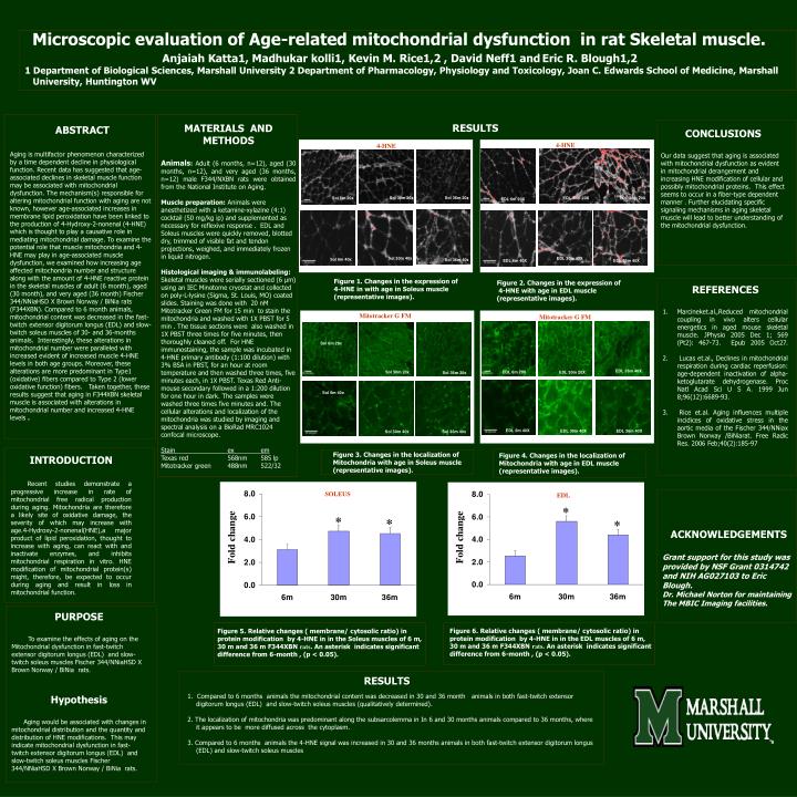
Figure 4. Changes in the localization of Mitochondria with age in EDL muscle (representative images). Figure 1. Changes in the expression of 4-HNE in with age in Soleus muscle (representative images). Figure 2. Changes in the expression of 4-HNE with age in EDL muscle (representative images). Microscopic evaluation of Age-related mitochondrial dysfunction in rat Skeletal muscle. Anjaiah Katta1, Madhukar kolli1, Kevin M. Rice1,2 , David Neff1 andEric R. Blough1,2 1 Department of Biological Sciences, Marshall University 2 Department of Pharmacology, Physiology and Toxicology, Joan C. Edwards School of Medicine, Marshall University, Huntington WV RESULTS MATERIALS AND METHODS Animals:Adult (6 months, n=12), aged (30 months, n=12), and very aged (36 months, n=12) male F344/NXBN rats were obtained from the National Institute on Aging. Muscle preparation: Animals were anesthetized with a ketamine-xylazine (4:1) cocktail (50 mg/kg ip) and supplemented as necessary for reflexive response . EDL and Soleus muscles were quickly removed, blotted dry, trimmed of visible fat and tendon projections, weighed, and immediately frozen in liquid nitrogen. Histological imaging & immunolabeling: Skeletal muscles were serially sectioned (6 μm) using an IEC Minotome cryostat and collected on poly-L-lysine (Sigma, St. Louis, MO) coated slides. Staining was done with 20 nM Mitotracker Green FM for 15 min to stain the mitochondria and washed with 1X PBST for 5 min . The tissue sections were also washed in 1X PBST three times for five minutes, then thoroughly cleaned off. For HNE immunostaining, the sample was incubated in 4-HNE primary antibody (1:100 dilution) with 3% BSA in PBST, for an hour at room temperature and then washed three times, five minutes each, in 1X PBST. Texas Red Anti-mouse secondary followed in a 1:200 dilution for one hour in dark. The samples were washed three times five minutes and. The cellular alterations and localization of the mitochondria was studied by imaging and spectral analysis on a BioRad MRC1024 confocal microscope. Stain ex em Texas red 568nm 585 lp Mitotracker green 488nm 522/32 ABSTRACT CONCLUSIONS Our data suggest that aging is associated with mitochondrial dysfunction as evident in mitochondrial derangement and increasing HNE modification of cellular and possibly mitochondrial proteins. This effect seems to occur in a fiber-type dependent manner . Further elucidating specific signaling mechanisms in aging skeletal muscle will lead to better understanding of the mitochondrial dysfunction. 4-HNE 4-HNE 4-HNE Aging is multifactor phenomenon characterized by a time dependent decline in physiological function. Recent data has suggested that age-associated declines in skeletal muscle function may be associated with mitochondrial dysfunction. The mechanism(s) responsible for altering mitochondrial function with aging are not known, however age-associated increases in membrane lipid peroxidation have been linked to the production of 4-Hydroxy-2-nonenal (4-HNE) which is thought to play a causative role in mediating mitochondrial damage. To examine the potential role that muscle mitochondria and 4-HNE may play in age-associated muscle dysfunction, we examined how increasing age affected mitochondria number and structure along with the amount of 4-HNE reactive protein in the skeletal muscles of adult (6 month), aged (30 month), and very aged (36 month) Fischer 344/NNiaHSD X Brown Norway / BiNia rats (F344XBN). Compared to 6 month animals, mitochondrial content was decreased in the fast-twitch extensor digitorum longus (EDL) and slow-twitch soleus muscles of 30- and 36-months animals. Interestingly, these alterations in mitochondrial number were paralleled with increased evident of increased muscle 4-HNE levels in both age groups. Moreover, these alterations are more predominant in Type1 (oxidative) fibers compared to Type 2 (lower oxidative function) fibers. Taken together, these results suggest that aging in F344XBN skeletal muscle is associated with alterations in mitochondrial number and increased 4-HNE levels . EDL 30m 20x EDL 36m 20x Sol 6m 20x Sol 30m 20x Sol 36m 20x EDL 30m 20X EDL 36m 20X Sol 6m 20x EDL 6m 20X EDL 6m 20X EDL 6m 40x EDL 30m 40x EDL 36m 40x Sol 6m 40x Sol 30m 40x EDL 30m 40X EDL 6m 40X Sol 6m 40x Sol 36m 40x EDL 6m 40X EDL 36m 40X • REFERENCES • Marcineket.al.,Reduced mitochondrial coupling in vivo alters cellular energetics in aged mouse skeletal muscle. JPhysio 2005 Dec 1; 569 (Pt2): 467-73. Epub 2005 Oct27. • Lucas et.al., Declines in mitochondrial respiration during cardiac reperfusion: age-dependent inactivation of alpha-ketoglutarate dehydrogenase. Proc Natl Acad Sci U S A. 1999 Jun 8;96(12):6689-93. • Rice et.al. Aging influences multiple incidices of oxidative stress in the aortic media of the Fischer 344/NNiax Brown Norway /BiNiarat. Free Radic Res. 2006 Feb;40(2):185-97 Mitotracker G FM Mitotracker G FM EDL 6m 20X Sol 6m 20x Sol 6m 20x Sol 36m 20x Sol 6m 20x EDL 6m 20x Sol 30m 20x EDL 30m 20x EDL 36m 20x EDL 36m 40X EDL 6m 20X EDL 36m 20X Sol 30m 20x EDL 30m 20X Sol 36m 20x Sol 6m 40x Sol 6m 40x EDL 6m 40x Sol 6m 40x Sol 30m 40x EDL 30m 40x Sol 36m 40x EDL 36m 40x EDL 6m 40X EDL 6m 40X EDL 30m 40X EDL 36m 40X Sol 30m 40x Sol 36m 40x Figure 3. Changes in the localization of Mitochondria with age in Soleus muscle (representative images). INTRODUCTION Recent studies demonstrate a progressive increase in rate of mitochondrial free radical production during aging. Mitochondria are therefore a likely site of oxidative damage, the severity of which may increase with age.4-Hydroxy-2-nonenal(HNE),a major product of lipid peroxidation, thought to increase with aging, can react with and inactivate enzymes, and inhibits mitochondrial respiration in vitro. HNE modification of mitochondrial protein(s) might, therefore, be expected to occur during aging and result in loss in mitochondrial function. SOLEUS EDL ACKNOWLEDGEMENTS Grant support for this study was provided by NSF Grant 0314742 and NIH AG027103 to Eric Blough. Dr. Michael Norton for maintaining The MBIC Imaging facilities. PURPOSE To examine the effects of aging on the Mitochondrial dysfunction in fast-twitch extensor digitorum longus (EDL) and slow-twitch soleus muscles Fischer 344/NNiaHSD X Brown Norway / BiNia rats. Hypothesis Aging would be associated with changes in mitochondrial distribution and the quantity and distribution of HNE modifications. This may indicate mitochondrial dysfunction in fast-twitch extensor digitorum longus (EDL) and slow-twitch soleus muscles Fischer 344/NNiaHSD X Brown Norway / BiNia rats. Figure 6. Relative changes ( membrane/ cytosolic ratio) in protein modification by 4-HNE in in the EDL muscles of 6 m, 30 m and 36 m F344XBN rats. An asterisk indicates significant difference from 6-month , (p < 0.05). Figure 5. Relative changes ( membrane/ cytosolic ratio) in protein modification by 4-HNE in in the Soleus muscles of 6 m, 30 m and 36 m F344XBN rats. An asterisk indicates significant difference from 6-month , (p < 0.05). • RESULTS • 1. Compared to 6 months animals the mitochondrial content was decreased in 30 and 36 month animals in both fast-twitch extensor digitorum longus (EDL) and slow-twitch soleus muscles (qualitatively determined). • 2. The localization of mitochondria was predominant along the subsarcolemma in In 6 and 30 months animals compared to 36 months, where it appears to be more diffused across the cytoplasm. • 3. Compared to 6 months animals the 4-HNE signal was increased in 30 and 36 months animals in both fast-twitch extensor digitorum longus (EDL)and slow-twitch soleus muscles