Download
1 / 39
390 likes | 583 Vues
Development and Sex Determination. Chapter 7. 7.1 The Human Reproductive System. The human reproductive system We will not cover most of this information in this course because it is focused on anatomy rather than human genetics. Timing of Meiosis and Gamete Formation in Males and Females.

E N D
Development and Sex Determination Chapter 7
7.1 The Human Reproductive System • The human reproductive system • We will not cover most of this information in this course because it is focused on anatomy rather than human genetics.
Timing of Meiosis and Gamete Formation in Males and Females • Males • Spermatogenesis begins during puberty • Millions of sperm are always in production • Spermatogenesis takes about 48 days • Each cell that undergoes meiosis produces 4 sperm • Females • Primary oocytes produced during embryonic development remain in meiosis I until ovulation • Ovulation begins during puberty • Meiotic division produces 1 large oocyteand 2-3 polar bodies
The Largest Cell • The human oocyte is the largest cell produced in the body. It is large enough to be seen with the naked eye
7.2 From Fertilization to Birth • Cell divisions in the zygote form an early embryonic stage called the blastocyst • Blastocyst • The developmental stage at which the embryo implants into the uterine wall • Stem cells are derived from a blastocyst • Inner cell mass • A cluster of cells in the blastocyst that gives rise to the fetus
Implantation • Implantation • The embryo implants in the uterine wall, and membranes develop to support the embryo • Trophoblast • Outer layer of cells in the blastocyst that gives rise to the membranes surrounding the embryo
Embryonic Membranes and Placenta • Chorion • formed from trophoblast • Releases human chorionic gonadotropin (hCG) hormone which maintains uterine lining and stimulates endometrial cells to produce hormones—hCG is what pregnancy tests detect • Grows to eventually form the placenta
Trophoblast (surface layer of cells of the blastocyst) Fertilization Endometrium Blastocoel Implantation Endometrium Inner cell mass Inner cell mass Uterine cavity 1 2 3 4 5 Fig. 7-6a, p. 156
Chorion Start of amniotic cavity Start of embryonic disk Chorionic cavity Chorionic villi Blood-filled spaces Amniotic cavity Start of yolk sac Connective tissue Start of chorionic cavity Yolk sac 8 7 6 Actual size Actual size Actual size Fig. 7-6b, p. 156
Development is Divided into Three Trimesters • First trimester • First month: basic tissue layers form; most of the body is divided into paired segments • Second month: most major organ systems are formed • Third month: embryo becomes a fetus; sexual development is initiated
Development is Divided into Three Trimesters • Second trimester • Increase in size and organ-system development • Bony parts of skeleton form • Heartbeat is heard with a stethoscope • Fetal movements begin • Third trimester • Rapid growth • Circulatory and respiratory systems mature • Birth is a hormonally induced process at the end of the 3rd trimester
WEEKS 5–6 Head growth exceeds growth of other regions Retinal pigment Future external ear Upper-limb differentiation (hand plates develop, then digital rays of future fingers; wrist, elbow start forming) Umbilical-cord formation between weeks 4 and 8 (amnion expands, forms tube that encloses the connecting stalk and a duct for blood vessels) Foot plate (b) Actual length Fig. 7-7ab, p. 158
WEEK 8 Final week of embryonic period; embryo looks distinctly human compared to other vertebrate embryos Upper and lower limbs well formed; fingers and then toes have separated Primordial tissues of all internal, external structures now developed Tail has become stubby (c) Actual length Fig. 7-7cd, p. 159
Placenta WEEK 16 Length: 16 centimeters (6.4 inches) 200 grams (7 ounces) Weight: WEEK 29 Length: 27.5 centimeters (11 inches) 1,300 grams (46 ounces) Weight: During fetal period, length measurement extends from crown to heel (for embryos, it is the longest measurable dimension, as from crown to rump). WEEK 38 (full term) Weight: 50 centimeters (20 inches) 3,400 grams (7.5 pounds) Length: (d) Fig. 7-7cd, p. 159
7.3 Teratogens Are a Risk to the Developing Fetus • Teratogen • Any physical or chemical agent with the potential to cause birth defects • Radiation, viruses, medications, alcohol
Alcohol is a Teratogen • Fetal alcohol syndrome (FAS) • A range of birth defects caused by maternal alcohol consumption during pregnancy • Alcohol is the most common teratogenic problem and leading cause of preventable birth defects • There is no “safe” amount of alcohol consumption during pregnancy
Tetatogens and their impact on organ formation Defects in physiology; physical abnormalities minor Major morphological abnormalities Weeks: 1 2 3 4 5 6 7 8 9 16 20–36 38 Cleavage, implantation Future heart Future eye Future ear Palate forming Future brain Limb buds External genitalia Teeth Central nervous system Heart Upper limbs Eyes Lower limbs Teeth Palate External genitalia Insensitivity to teratogens Ear Fig. 7-8, p. 160
Mechanisms of Sex Determination Mechanisms of sex determination vary from species to species XX/XY system XX/X0 ZW/ZZ Temp.
Human Sex Ratios • Sex ratio • The proportion of males to females changes throughout the life cycle • The ratio at conception is slightly higher for males. (**prenatal deaths most likely due to lethal X-linked recessive alleles) • The ratio at birth is about 105 males/100 females • The ratio of females to males increases as a population ages
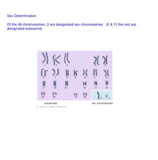
7.5 Defining Sex in Stages: Chromosomes, Gonads, and Hormones • Sex of an individual is defined at three levels • Chromosomal sex (established at fertilization) • Gonadalsex (begins around week 7 or 8 of embryogenesis) • Phenotypic sex • Gonadal and phenotypic sex depend on the interaction of genes and environmental factors, especially hormones
Gonadal Sex Differentiation • For the fist 7 or 8 weeks, the embryo is neither male nor female • Both male and female reproductive duct systems begin to develop • Genes cause gonads to develop as testes or ovaries, establishing gonadal sex • Alternative pathways produce an intermediate sex for 1 out of every 2000 births.
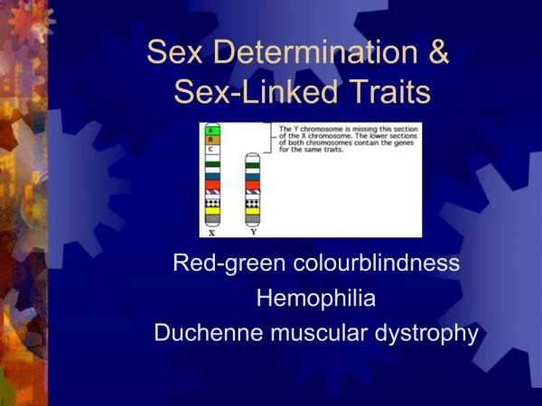
Y Chromosome and Testis Development • SRY gene • Sex-determining region of the Y chromosome • Plays a major role in causing the undifferentiated gonad to develop into a testis • Testis development causes secretion of testosterone • Müllerian inhibiting hormone (MIH) • Hormone produced by developing testis that causes breakdown of Müllerian (female) ducts in the embryo
Female Development • Requires the absence of the Y chromosome and the presence of two X chromosomes for the embryonic gonad to develop as an ovary • In the absence of testosterone, the Wolffian duct system degenerates • In the absence of MIH, the Müllerian duct system forms female reproductive system
Egg with X sex chromosome Male Female Fertilized by Fertilized by Sperm with X chromosome Sperm with Y chromosome Chromosomal sex Embryo with XX sex chromosomes Embryo with XY sex chromosomes Sex-determining region of the Y chromosome (SRY) brings about development of undifferentiated gonads into testes. No Y chromosome, so no SRY. With no masculinizing influence, undifferentiated gonads develop into ovaries. Gonadal sex Testes secrete masculinizing hormones, including testosterone, a potent androgen. No androgens secreted In presence of testicular hormones, undifferentiated reproductive tract and external genitalia develop along male lines. With no masculinizing hormones, undifferentiated reproductive tract and external genitalia develop along female lines. Phenotypic sex Fig. 7-14, p. 167
Androgen Insensitivity • Androgen insensitivity (CAIS) • A mutation in the X-linked androgen receptor gene (AR) causes XY males to become phenotypic females • Testosterone is produced, but not testosterone receptors; cells develop as females
XY Female with Androgen Insensitivity Fig. 7-15, p. 168
Exploring Genetics: Joan of Arc or John of Arc? • Joan of Arc fought with the French at the Battle of Orleans, and was burned as a heretic by her enemies, the English, in 1431 • From an examination of trial evidence and records of her physical examinations, R.B. Greenblatt proposed that Joan had phenotypic characteristics of androgen insensitivity
Mutations can cause Sex Phenotypes to Change at Puberty • Pseudohermaphroditism • Mutations in several different genes cause XY individuals to develop the phenotype of females • Affected individuals have structures that appear female at birth • At puberty, testosterone burst causes a change into the male phenotype
7.7 Equalizing the Expression of X Chromosomes in Males and Females • Lyon hypothesis (proposed by Mary Lyon) • How do females avoid getting a double dose of protein from X-linked genes? • Random inactivation of one X chromosome in females equalizes the activity of X-linked genes • Barr body • A densely staining mass in the somatic nuclei of mammalian females • An inactivated X chromosome, tightly coiled
X Chromosomes and Barr Bodies Fig. 7-16, p. 169
Female Mammals are Mosaics for X Chromosome Expression • In females, some cells express the mother’s X chromosome and some cells express the father’s X chromosome • Inactivated chromosome can come from either mother or father • Inactivation occurs early in development • Inactivation is permanent; all descendants of a particular cell have the same X inactivated
Female Mammals are Actually Mosaics for X Chromosome Expression
Female Mammals are Mosaics for X Chromosome Expression Unaffected skin (X chromosome with recessive allele was condensed; its allele is inactivated. The dominant allele on other X chromosome is being expressed in this tissue.) Affected skin with no normal sweat glands (yellow). In this tissue, the X chromosome with dominant allele has been condensed. The recessive allele on the other X chromosome is being transcribed. (a) (b) Fig. 7-18, p. 171
Mosaic Expression in Female Mammals The gene for fur color in cats is on the X chromosome. Fig. 7-17, p. 170
Inactivation of X Chromosome by XIST RNA XIST gene codes for RNA that binds to the X chromosome and inactivates it Fig. 7-19, p. 171
7.8 Sex-Related Phenotypic Effects • Sex-limited trait - affects a structure or function of the body that is present in only males or females • Women do not get prostate cancer, women do not grow beards but pass on the gene for beard growth on to their sons • Sex-influenced trait - an allele is dominant in one sex and recessive in the other • Baldness—the allele is dominant in males and recessive in females
Imprinting • Imprinting • difference in expression of a gene depending upon whether it was inherited from mother or father • More discussion to follow on this topic in Chapter 11.