Download
1 / 60
620 likes | 665 Vues
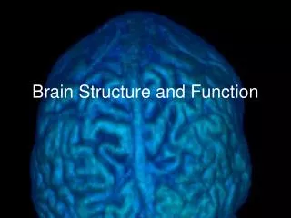
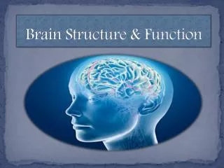
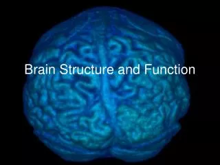
Gross Structure of the Brain. Won Taek Lee, M.D., Ph.D. Deptartment of Anatomy Yonsei Univ. College of Medicine. Nervous System. CNS (central nervous system) Brain Spinal Cord PNS (peripheral nervous system) Peripheral Nerve Ganglion.

E N D
Gross Structure of the Brain Won Taek Lee, M.D., Ph.D. Deptartment of Anatomy Yonsei Univ. College of Medicine
Nervous System CNS (central nervous system) Brain Spinal Cord PNS (peripheral nervous system) Peripheral Nerve Ganglion
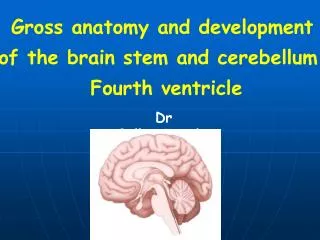
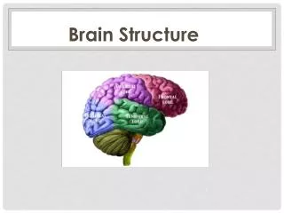
Gross Divisions of the Brain Cerebrum (cerebral hemisphere) Diencephalon Cerebellum Brainstem
Primary vesicle Secondary vesicle Derivatives Prosencephalon telencephalon Cerebral cortex Cerebral white matter Basal ganglia diencephalon Thalamus Hypothalamus Subthalamus Epithalamus Mesencephalon mesencephalon Midbrain Rhombencephalon metencephalon Cerebellum Pons myelencephalon Medulla oblongata Embryonic (developmental) divisions of the Brain
Inner Structures of the Brain • Basal ganglia • Thalamus • Hypothalamus • Midbrain • Pons • Medulla oblongata
Cerebrum – Cerebral Hemisphere Telencephalic structure –lateral ventricles • Cerebral Cortex • - lobes, gyrus & sulcus • 2. Cerebral White Matter • (medullary center) • 3. Basal Ganglia
Lobes of the Cerebral Cortex (lateral surface) • Frontal lobe • Parietal lobe • Temporal Lobe • Occipital lobe • Limbic lobe
Lobes of the Cerebral Cortex (medial surface) • Frontal lobe • Parietal lobe • Temporal Lobe • Occipital lobe • Limbic lobe
Lobes of the Cerebral Cortex (basal surface) • Frontal lobe • Parietal lobe • Temporal Lobe • Occipital lobe • Limbic lobe
Gyri of the Frontal Lobe Lateral Surface Precentral Gyrus --- primary motor area Superior Frontal Gyrus Middle Frontal Gyrus Inferior Frontal Gyrus Pars Opercularis Pars Triangularis Pars Orbitalis Medial Surface Medial Frontal Gyrus Paracentral Lobule Basal Surface Rectus Gyrus Orbital Gyrus Inferior Frontal Gyrus ]Broca’s area(dominant hemisphere)
Gyri of the Cerebral Cortex (Lateral Surface) Broca’s area Pars Opercularis Pars Triangularis Pars Orbitalis
Broca’s Area Pars triangularis and pars opercularis of the inferior frontal gyrus of dominant hemisphere. Photograph of the brain of Broca’s patient called “Tan” (real name is Leborgne).
Gyri of the Cerebral Cortex (basal surface)
Gyri of the Parietal Lobe Lateral Surface Postcentral Gyrus ---- primary somesthetic area Superior Parietal Lobule Inferior Parietal Lobule ---- Wernicke’s area Supramarginal Gyrus Angular Gyrus Medial Surface Paracentral Lobule Precuneus
Gyri of the Occipital Lobe Lateral Surface Lateral Occipital Gyrus Superior Occipital Gyrus Inferior Occipital Gyrus Medial Surface Cuneus Lingual Gyrus Basal Surface Lingual Gyrus Occipitotemporal Gyrus
Gyri of the Cerebral Cortex (basal surface)
Gyri of the Temporal Lobe Lateral Surface Superior Temporal Gyrus Middle Temporal Gyrus Inferior Temporal Gyrus Basal Surface Lingual Gyrus Occipitotemporal Gyrus Medial Occipitotemporal Gyrus Lateral Occipitotemporal Gyrus
Gyri of the Cerebral Cortex (basal surface)
Gyri of the Limbic Lobe Outer Ring Cingulate Gyrus Isthmus of Cingulate Gyrus Parahippocampal Gyrus Uncus, Uncinate Gyrus Inner Ring Hippocampal Formation Hippocampus, Ammon's Horn Dentate Gyrus Fasciolar Gyrus Indusium Griseum (Supracallosal Gyrus) Paraterminal Gyrus (Subcallosal Gyrus) Subcallosal Area (Parolfactory Area)
Gyri of the Cerebral Cortex (basal surface)
1 2 5 4 3 Structures of the Lateral Surface of the Brain
3 2 4 1 Structures of the Medial Surface of the Brain
2 1 Structures of the Basal Surface of the Brain
Structures of Fornix • Uncus • Hippocampus • Fimbria • Crus • Commissure • Corpus • Column • Mammillary body 6 7 5 8 3 2 1 4
Cerebral White Matter Projection fiber Corona radiata Commissural fiber Corpus callosum rostrum, genu, trunk, splenium Anterior commissure Commissure of fornix Association fiber Short association fiber Long association fiber
Fibers of Cerebral White Matter (coronal section) 1. Corpus callosum 2. Internal capsule 3. Superior occipitofrontal fasciculus 4. Superior longitudinal fasciculus 5. Inferior occipitofrontal fasciculus 6. Cingulum 7. Uncinate fasciculus 8. Inferior longitudinal fasciculus
Basal Ganglia (traditional concept) Corpus striatum lenticular nucleus putamen globus pallidus caudate nucleus Amygdaloid body
Basal Ganglia 3 2 1 3 2 1 4
Basal Ganglia 2 1 1 2 3 3
Diencephalon Thalamus Hypothalamus Epithalamus Subthalamus 3rd ventricle and tela choroidea
Diencephalon 1 4 5 2 1 2 3 3
Ventral Surface of Diencephalon 4 1 5 5 2 3 6
Cerebellum Anterior Lobe Posterior Lobe Flocculonodular Lobe flocculus, nodulus Vermis Paravermal Region Cerebellar Hemisphere