Download

1 / 43
430 likes | 542 Vues
The Biochemistry of Cell Death Pál Bauer 04.30.2009. CELL PROLIFERATION. Tissue Turnover(days) Daily proliferation Gut-Jejunum 1-2 50-100% Bone Marrow 2-4 25-50% Thymus 7 14%

E N D
CELL PROLIFERATION • Tissue Turnover(days) Daily proliferation • Gut-Jejunum 1-2 50-100% • Bone Marrow 2-4 25-50% • Thymus 7 14% • Bladder 30-60 2-3% • Liver 450 0.2% • Endothelial Cells >500 <0.2% • Brain >800 <0.1%
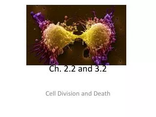
Types of cell death Necrosis – non-apoptotic accidental cell death (common definition) <or> morphology seen after a cell has already died (pathology; has nothing to do with biochemistry of how the cell died) Autophagy – degradation of cellular components within dying cells in an autophagic vacuole; begins with sequestration of cytoplasmic material within phagosomes, under control of GTPases and phosphatidylinositol kinases Oncosis – prelethal pathway leading to cell death; accompanied by cellular swelling, organelle swelling, blebbing, and increased membrane permeability; oncotic cells proceed to necrosis with lysis and spillage of contents before being recognized by phagocytosis; inflammation results Apoptosis…..
Apoptosis Necrosis Tightly regulated and controlled Not regulated or controlled Active participation of cellular components Passive process Follows a specific ordered pattern of events Cell swells and disintegrates in a disordered manner No leakage of cellular contents Rupture of cell membrane results in the leakage of cellular contents into extracellular space No inflammation Associated with inflammation Induced by cell signaling or slight damage to the cell Induced by massive cellular injury Kerr, Wyllie, and Currie, Brit. J. Cancer 26:239, 1972
Importance of Apoptosis • Maintaining homeostasis • Cell death is balanced with mitosis to regulate • total cell number • Embryonic development • Errors in apoptosis can lead to birth defects • Improper regulation contributes to human disease
Apoptosis vs Autophagy vs Necrosis: Three deaths or one continuum Cells are genetically programmed to initiate cell death upon a) exposure to a death ligand (death by design) or b) removal of factors required for cell survival (death by default). Suicide Hallmarks: Cell is disassembled through a series of orderly events orchestrated by gene expression and needs ATP
Apoptosis vs Autophagy vs Necrosis: Three deaths or one continuum If cellular homeostasis is severely compromised, a cell cannot maintain a stable internal environment and is lysed. Environmental conditions that trigger necrosis include acutelack of oxygen, elevated temperature, contact with toxic compounds, excessive mechanical strain (trauma). Murder Hallmarks: Cell explodes. Cytoplasmic contents are released eliciting an inflammatory response
Apoptosis vs Autophagy vs Necrosis: Three deaths or one continuum When cells are no longer exposed to sufficient nutrients in their microenvironment, they can cannibalize some of their internal organelles such as the mitochondria to re-use these components. A catabolic process by which cells degrade and digest their own cytoplasmic constituents, usually through the action of lysosomal enzymes. One of the most distinguishing features of autophagy is the dynamic rearrangement of cellular membrane to sequester cytosol and organelles into autophagosomes for delivery to the lysosome or vacuole. Self-cannibalism Autophagy Hallmarks: Formation of a double membrane within the cell which envelops the materials to be degraded into a vesicle called an autophagosome. The autophagosome then fuses with a lysosome whose hydrolytic enzymes degrade the materials
Cellular changes associated with apoptosis: Early events Various apoptotic signals cause outer mitochondrial membrane to become permeable Cells round up and lose contact with neighboring cells and ECM Loss of lipid asymmetry in plasma membrane
Cellular changes associated with apoptosis: DNA Nucleus becomes convoluted and buds into several fragments Apoptotic cell Normal cell
Chromatin aggregates into dense compact masses DNA is digested by endonucleases Normal cell Apoptotic cell Cellular changes associated with apoptosis: DNA
Plasma membrane blebs Cell breaks up into membrane spheres (“apoptotic bodies”) Phagocytic cells remove apoptotic bodies Cellular changes associated with apoptosis: Terminal events
No. of cells G1 G2 S DNA content Methods
(Riedl & Shi. (2004). Nat. Rev. Mol. Cell. Biol.5: 897). Conservation of the Apoptotic Pathway
Caspases Cysteine-dependent aspartate specific proteases Have a cysteine at the active site Cleave target just after aspartic acid residues Substrate specificity is determined by the 4 residues upstream of cleavage site Exist in cytosol as single chain proenzymes (procaspases) which are activated when cleaved by other caspases Once activated, cleave other procaspases – results in proteolytic cascade Also cleave key proteins in the cell, causing the characteristic morphology and biochemistry of apoptosis.
Caspases • Cysteine proteases that cleave after an aspartate • At least 14 mammalian caspases (11 human) • At least 7 of the 14 are important for apoptosis. • Caspases are made as proteolytically inactive zymogens/proenzymes • Removal of the propeptide not necessary Caspases (Riedl & Shi. (2004). Nat. Rev. Mol. Cell. Biol.5: 897).
Initiator Caspases • caspase-2, -8, -9 and –10 • Extended N-terminal region with one or more adaptor domains. • Auto-activated • Regulated by protein interactions • Effector Caspases • Caspase-3, -6, -7 • Contain 20-30 residue-long propeptides • Activated by proteolytic cleavage by initiator caspases. (Riedl & Shi. (2004). Nat. Rev. Mol. Cell. Biol.5: 897). Black arrows: Activation of enzyme activity by proteolytic cleavate between p20 and p10. Grey arrows: Modulation of enzyme activity by proteolytic cleavage (not mediated by caspases) CARD: Caspase-recruitment domain. DED: Death-effector domain. L1-4: Loops forming the catalytic site. The 2 Classes of Caspases
(Riedl & Shi. (2004). Nat. Rev. Mol. Cell. Biol.5: 897). Activation of Caspase Enzyme Activity • Caspases are activated by cleavage of the L2-loop • Conformational changes of L1-4 • Additional regulation of caspase activity: • Transcriptional • Posttranslational modifications. • Inhibitor-of-apoptosis (IAP) proteins • Proteasome-mediated removal
Fas – Fas ligand system- Extrinsic pathway Fas Prototypical cell death receptor of the TNF receptor superfamily No intrinsic enzymatic activity Signals via adaptor proteins Fas ligand (FasL) Only expressed by activated T cells Transmembrane TNF-like protein When the TCR (T cell receptor) of an antigen specific CTL (cytotoxic T lymphocyte) binds antigen on MHC I, the expression of FasL is induced on the T cell FasL binds Fas (present on most cells of body) that is on the presenting cell to induce death of that cell Cytoplasmic tail of Fas binds its adaptor protein FADD (Fas Associated Death Domain protein) Fas-FADD complex binds to and activates caspase 8 Initiates lethal proteolytic cascade
(Curtin & Cotter (2003). Cell. Signalling. 15: 983). ”Death Ligands” and ”Death Receptors”
Intrinsic Pathway Usually initiated by cellular stress (UV, cytotoxic drugs etc.) usually causing alterations in mitochondria membrane potential (MMP). Mitochondria-dependent: mitochondria sequester pro-apoptotic proteins, e.g. cytochrome C. Regulated by Bcl-2 family member of proteins – regulate the release of pro-apoptotic factors from mitochondria. Changes in MMP free cytochrome c from intermembrane space out into the cytosol Cytochrome c then combines with dATP, APAF-1 (apoptotic protease activating factor-1), and procaspase 9 to form a catalytic complex called the apoptosome (Caspase 9 co-factor is APAF-1 which must be bound by cytochrome c to drive Caspase 9 into its active conformation) Apoptosome activates caspases 3 and 7 (effector caspases) Effector caspases Nature407, 770-776 (12 October 2000)
Regulation of intrinsic pathway Bcl-2 family of proteins (gene orig. isolated from a B-cell lymphoma): Pro apoptotic effects: e.g. Bax (indirectly regulated by tumor repressor P53), Bad, and Bak Anti apoptotic effects: e.g. Bcl-2, Bcl-XL These proteins may be localized to mitochondria intermembrane or targeted to the organelle in response to stimulus Regulate release of pro-apoptotic factors from mitochondria, especially cytochrome c. Also release Smac (second mitochondria-derived activator of caspases) and DIABLO (direct IAP-binding protein with low pI) which bind to IAPs (Inhibioros of Apoptosis proteins) Battle between pro and anti levels to determine cell’s response to apoptotic stimuli such as ROS (reactive oxygen species), Ca++, radiation etc. Also released from mitochondria is AIF – apoptosis inducing factor; once released, it translocates to nucleus to induce chromatin condensation and DNA fragmentation
How do proteins get released from mitochondria? Several hypotheses for how Bcl-2 proteins regulate release of cytochrome C: By forming channels in mitochondria membrane By interacting with other proteins to form channels By inducing rupture of the mitochondrial membrane By oligomerizing to form a weakly selective ion channel. BcL-2 family proteins regulate the release of apoptogenic cytochrome c by the mitochondrial channel VDAC (voltage dependent anion channel) Nature 399, 483-487 (1999))
The cytochrome c-induced caspase activation pathway Inactive form Conformation change oligomerization
Differences between pathways Mitochondrial • Default pathway • Stress mediated • Requires new protein synthesis • Takes 12-24 hours to occur Death Domain • Instructive apoptosis • Protein synthesis not required (blocking protein synthesis accelerates death) • Very rapid – death in a few hrs.
Effector Caspases Activated effector caspases cleave target proteins: Nuclear Lamins – scaffold proteins of nuclear envelope; leads to nuclear shrinkage and fragmentation Cytoskeleton proteins – e.g.: Fodrin; leads to loss of cell shape and membrane blebbing; Gelsolin (an actin depolymerizing enzyme); cleaved by caspase 3; role in cell morphology during apoptosis (blebbing etc.) ICAD (inhibitor of Caspase Activated Dnase) – DNA now cut up by CAD Components of focal adhesion complex; leads to detachment of apoptotic cells from other cells Other caspase dependent features: Cleavage of PAK2 (member of p21-activated kinase family); results in formation of apoptotic bodies Exposure of phosphatidylserine on outer membrane; probably due to down-regulation of phospholipid translocase activity and/or activation of lipid scramblase
INHIBITORS of APOPTOSIS • Bcl2 family members • Decoy receptors • Inhibitors of apoptotic signal transfer • Caspase inhibitors - viral origin, crmA,p35 • - IAP,NAIP, survivin • - small molecular weight inhibitors
(Chan & Yu. (2004). Clin. Exp. Pharmacol. Physiol. 31: 119). The Bcl-2 Family • BH1, BH2: Predicted to form ion channels • BH3: The ”suicide domain” – regulates cell death • BH4: Thought to confer anti-apoptotic activity. • The Bcl-2 Family • Proteins with anti- or pro-apoptotic function • Characteristics: • (Often) a C-terminal transmembrane domain. • Bcl-2 homology (BH) domains. • Bcl-2 proteins form homo- and heterodimers via their BH-domains
Rheostat model by SJ Korsmeyer et al. Relative concentration of bcl-2/Bax family member interactions determines apoptotic fate of cell. In the presence of excess of Bax, Bcl-2 is displaced from Apaf-1, allowing the activation and cleavage of caspase, and mediate the release of cyto c from the mitochondria. Cell79: 189-192, 1994
Corpse clearing A defining point of apoptosis is to clear the dying cell before it can release inflammatory molecules Macrophages ingesting apoptotic cells release anti-inflammatory and immunosuppressive cytokine transforming growth factor-beta1 (TGF-β1) Macrophages ingesting necrotic cells will release pro-inflammatory mediators Cells undergoing apoptosis show changes in surface of plasma membrane. These “eat me” signals are recognized by phagocytes Exposure of phosphatidyl serine (PS) – PS receptor is expressed on phagocytes Change in cell surface sugars – detected by lectins on phagocyte cell Sites that bind “bridging molecules” e.g. C1q – C1q deficiency leads to impaired phagocytosis of apoptotic cells ICAM-3 – binds alternate receptor on macrophages; possibly CD14 Macrophages thought to “tether” dying cells by using CD14 or beta integrin before engaging receptors that drive apoptosis
PI3K/AKT pathway PIP2 PI3K Src PTEN Proliferation Survival PIP3 PDK NF-kB IKK Akt Caspase MDM2 FOXO mTOR p27 p21 BAD ASK GSK3b p70 S6K Bcl-2 FasL b-catenin p53 4EBP protein translation Apoptosis Resistance Cell Cycle Progression proliferation Migration
Apoptotic cell Normal cell Methods Caspase activity: Activity assays Antibodies against active Cytochrome c Detection of release of cytochrome c from mitochondria to cytosol Mitochondria Detecting chages of Mitochondrial transmembrane potential DNA breakage TUNEL (Terminal deoxynucleotidyl Transferase-dUTP Nick End Labeling). TUNEL-staining Mitochondrial Transmembrane Potential Caspase Activity Red = active caspases Bright Green = DNA fragmentation (http://www.b-bridge.com/eng/products/apop/cdk.htm) (J. Biol. Chem.276:35891-9, 2001).