Download
1 / 26
260 likes | 380 Vues
Modeling the Cell Cycle Engine of Eukaryotes. John J. Tyson & Bela Novak Virginia Polytechnic Institute & State Univ. Budapest Univ. Technology & Economics.

E N D
Modeling the Cell CycleEngine of Eukaryotes John J. Tyson & Bela Novak Virginia Polytechnic Institute & State Univ. Budapest Univ. Technology & Economics
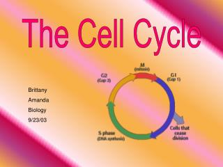
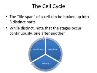
The cell cycle is the sequence of events by which a growing cell replicates all its components and divides them more-or-less evenly between two daughter cells... …so that the two daughter cells contain all the information and machinery necessary to repeat the process.
G1 cell division S (DNA synthesis) M (mitosis) G2
G1 Too small? DNA damage? G1/S checkpoint cell division S 1. Alternation of S and M phases (DNA synthesis) Unaligned chromosomes? 2. Balanced growth and division Metaphase checkpoint Unreplicated DNA? Too small? M (mitosis) G2 G2/M checkpoint
Cdk1 CycB Tar Tar- P G1 cell division Cyclin-dependent kinase S DNA replication M (mitosis) G2
Cdk1 CycB G1 G1/S cell division S DNA replication Exit M (mitosis) G2 G2/M
Wee1-P Wee1 Cdk1 Cdk1 CycB CycB P- less active Cdc25-P Cdc25 cyclin B degradation cyclin B synthesis less active active MPF cyclin B degradation less active
Cyclin Cdk1 Cyclin M Solomon’s protocol for cyclin-induced activation of MPF Ca2+ centrifuge Wee1 Cyclo- heximide Cdk1 Cdc25 cytoplasmic extract no synthesis of cyclin pellet no degradation of cyclin
Threshold MPF Cyclin (nM) Solomon et al. (1990) Cell 63:1013.
Frog egg Novak & Tyson (1993) J. Cell Sci.106:1153 active MPF no synthesis or degradation of cyclin total cyclin
hysteretic non-hysteretic MPF activity MPF activity Ti Ta T cyclin level cyclin level Prediction: The threshold concentration of cyclin B required to activate MPF is higher than the threshold concentration required to inactivate MPF.
Norel & Agur (1991). “A model for the adjustment of the mitotic clock by cyclin and MPF levels,” Science251:1076-1078. Tyson (1991). “Modeling the cell division cycle: cdc2 and cyclin interactions,” PNAS88:7328-7332. Goldbeter (1991). “A minimal cascade model for the mitotic oscillator involving cyclin and cdc2 kinase,” PNAS88:9107-9111. Novak & Tyson (1993). “Numerical analysis of a comprehensive model of M-phase control in Xenopus oocyte extracts and intact embryos,” J. Cell Sci.106:1153-1168. Thron (1996). “A model for a bistable biochemical trigger of mitosis,” Biophys. Chem.57:239-251. Thron (1997). “Bistable biochemical switching and the control of the events of the cell cycle,” Oncogene15:317-325.
G1 Start cell division S DNA replication Finish M (mitosis) G2 G2/M
CKI Cdh1 APC Cdc20 APC G1 Start cell division Cln2 S Clb5 DNA replication Cdk Finish Clb2 M (mitosis) G2 G2/M
Cdc20 P Cdk Cln2 P Cdh1 CKI AA Cdk Cdk AA CycB Cdk CycB Cdh1 Cdc14 Cdk CKI CycB CKI Cdc14
synthesis synthesis binding degradation degradation inactivation activation The mathematical model
G1 S/M Simulation of the budding yeast cell cycle mass CKI Cln2 Clb2 Cdh1 Cdc20 Time (min)
These are the “brutes” Kathy Chen Laurence Calzone 30 equations 100 parameters fitted by brute force
“With four parameters I can fit an elephant…” Is the model yeast-shaped?
k1 = 0.0013, v2’ = 0.001, v2” = 0.17, k3’ = 0.02, k3” = 0.85, k4’ = 0.01, k4” = 0.9, J3 = 0.01, J4 = 0.01, k9 = 0.38, k10 = 0.2, k5’ = 0.005, k5” = 2.4, J5 = 0.5, k6 = 0.33, k7 = 2.2, J7 = 0.05, k8 = 0.2, J8 = 0.05, … Parameter values Differential equations
Cdk Cln CKI CKI +APC Cdh1 +APC Cdk Cdc20 Cln Cdk CycB
Cdc14 Cln2 Mutual antagonism and bistability... Cdk CycB CKI Cdh1
Start Finish A/B Cdc14 Cln2 time S/G2/M Clb2/Cdk activity G1 A + Cln2 B+Cdc14
From molecular networks to cell physiology… 1.0 0.8 MPF 0.6 0.4 0.2 0 0 10 20 30 time (min) differential equations simulation & analysis Wee1 P Wee1 P Cdc2 Cdc2 CycB CycB Cdc25 P G2/M Cdc25 ??? molecules physiology
Our thanks to... National Science Foundation (USA) National Science Foundation (Hungary) National Institutes of Health James S. McDonnell Foundation Defense Advanced Research Project Agency