Download
1 / 61
630 likes | 937 Vues
TMJ, Face, Skull. TMJ. Mandibular fossa of temporal bone with condyle of mandible Incongruent surfaces Two joint cavities with articular disc interposed Lower cavity = hinge joint Upper joint = gliding. TMJ. TMJ. Mandible. Mandible. TMJ. Capsule Surrounds the joint Encloses the disc

E N D
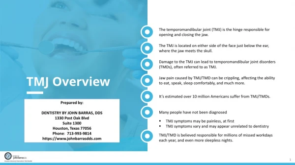
TMJ • Mandibular fossa of temporal bone with condyle of mandible • Incongruent surfaces • Two joint cavities with articular disc interposed • Lower cavity = hinge joint • Upper joint = gliding
TMJ • Capsule • Surrounds the joint • Encloses the disc • Attaches above the margins of the mandibular fossa • To the neck of the mandible • Inner aspect of capsule attaches to disc • Above disc – capsule loose • Below disc - taut
TMJ • Ligaments • Lateral ligament - AKA TMJ ligament • From zygomatic bone to run inferiorly and posteriorly to blend with the joint capsule to attach to lateral and posterior parts of the neck of the mandible • Sphenomandibular • Strong thin flat band lying on medial aspect of the joint • Passes inferiorly and forwards from the spine of the sphenoid to the lingula • Stylomandibular • Extends from the apex of the styloid process to the lower part of the posterior border of the ramus of the mandible, near the angle
TMJ • Innervated by CN V, Mandibular branch • Movements • Elevation, depression, retraction, protraction, side to side • Elevation and depression involves the hinge like rotation of the condyle against the disc in the lower compartment • Protraction and retraction – actions whereby the condyle and disc move as one unit against the mandibular fossa. In protraction the condyle and disc glide forwards so that the condyle rides on the articular eminence – retraction = opposite
TMJ • Side to side – grinding movements • Mandible is alternately protracted and retracted with the two sides moving in opposite directions so that one side is protracted while the other is retracted • Actions combined with elevation and depression, rhythmically and alternately
Muscles of Mastication • Masseter • Temporalis • Lateral pterygoid • Medial pterygoid • All innervated by CNV • Opening of jaw (depression) primarily passive or gravity assist
Nerve Supply to Face • Sensory by three divisions of CN V – opthalmic, maxillary, mandibular • Innervation of muscles of facial carried out by CN VII – the Facial Nerve • Origin, branches, motor functions, sensory functions, parasympatheric functions
Scalp • Three Layers • Outer = skin • Beneath that – subcutaneous layer with many nerves and vessels running through here, binds skin to inner layer • Galea Aponeurotica – AKA epicranial aponeurosis • Galea attaches to pericranium via loose CT • This allows scalp to move over the skull • Most muscles of face attach to skin, this arrangement allows them to be more mobile.
CN VII – The Facial Nerve • Motor nerve to muscles of facial expression with one notable exception • Origin = lower pons • Branches – common nerve enters face • Temporal • Zygomatic • Buccal • Mandibular • Cervical
CN VII • Motor Functions • Muscles of facial expression • External ear • Sensory functions • Ant. 2/3 of tongue • Soft palate • Pharynx • Parasympathetic • Gland stimulation
Muscles of Facial Expression • Primary action is to act as either a sphincter or dilator of the orifices of the face • Facial expression is a by-product • Orifices • Lips = labia • Nose = nares; Nostrils, Septum, Ala, Apex, Root • Eyelids = palpebrae • External Ear = auricle, lobule = soft portion
Selected Muscles of Facial Expression * = learn • Orbicularis Oculi • Levator Palpebrae Superioris* • O: Root of Orbital Cavity • I: Skin of upper eyelid • A: Raises upper eyelid • N: Note Well, Nerve = CN III • Ptosis is a condition of denervation to this muscle causing drooping of the eyelid, a clinical symptom indicating more loss
Muscles Continued • Occipitofrontalis • Corrugator • Auricular muscles of the ear – ant., post., sup. • Nose • Nasalis • Procerus • Depressor Septi
Muscles • Mouth • Depressor anguli oris • Depressor labii inferior • Mentalis • Risorius • Orbicularis oris • Buccinator* • Zygomaticus major • Zygomaticus minor • Levator labii superioris • Platysma