Download
1 / 20
200 likes | 334 Vues
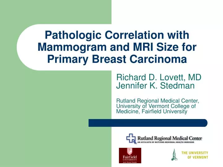
Pathologic Correlation with Mammogram and MRI Size for Primary Breast Carcinoma. Richard D. Lovett, MD Jennifer K. Stedman Rutland Regional Medical Center, University of Vermont College of Medicine, Fairfield University. Purpose.

E N D
Pathologic Correlation with Mammogram and MRI Size for Primary Breast Carcinoma Richard D. Lovett, MD Jennifer K. Stedman Rutland Regional Medical Center, University of Vermont College of Medicine, Fairfield University
Purpose • Current breast cancer screening practice is to employ physical examination, self examination, mammography • Magnetic Resonance Imaging (MRI) with contrast enhancement was demonstrated to be effective in 1989 for breast imaging by Kaiser and Heywang
Purpose • According to Bagley in 2003: • MRI accurately estimated the size of the breast lesion 91% of the time, an accuracy level believed to be greater than conventional mammograghy
Purpose • To examine all women at RRMC, who underwent MRI preoperatively, to determine whether the Mammogram or the MRI more accurately determined the presence and size of the pathologic lesion. The time frame used was 2001 until May 2006
138 0 138 152 56 51 27 10 4 4 Patients Male Female Lesions Pathologic Size 0-1cm >1-2cm >2-3cm >3-4cm >4-5cm >5cm Materials and Methods
Stage T1 (0-2cm) T2 (>2cm-5cm) T3 (>5cm) 107 (70%) 41 (27%) 4 (3%) Materials and Methods
Materials and Methods • Women with mammogram and MRI done preoperatively from 2001 to 5/2006 • Mammogram done by ACR accredited facility • MRI done at RRMC • Gadolinium enhancement • Dynamic contrast enhancement curves • Analysis of size and lesion by radiologist or radiation oncologist
Materials and Methods • Contrast enhanced MRI with breast cancer in the outer quadrant of the left breast • Images made with the subtracted gradient echo technique rather than the fat saturation technique
Materials and Methods • Dynamic enhancement curves plotted on the breast cancer with data points at 1 minute through 7 minutes after gadolinium injection
Materials and Methods • Patient with multifocal breast cancer in the right breast (the full extent of the breast cancer was not appreciated on the mammogram due to the patients young age (44)
Materials and Methods • Enhancement curves of many of the multifocal sites on this young woman • This patient decided on a mastectomy based on this data
Results- Pathologic Stage vs. Discordance Between Image and Pathology
Results r =0.44 r =0.64
Conclusions • The accuracy rate of MRI for predicting the size of the lesion is higher than the accuracy rate of mammogram 86% vs. 56% respectively • This finding is maintained for T1 and T2 lesions when staging is stratified • For T3 lesions, MRI is more accurate in predicting the size of the lesion, 25% to 0, however the small number of these lesions in the study precludes conclusions.
Conclusions • Mammography is more likely to image the breast cancer smaller than the true pathologic size • MRI with its contrast enhancement is more likely to image the lesion slightly larger than the pathologic size, presumed to be due to recruitment of perilesional blood vessels
Conclusions • Because the size of a lesion is one of the factors considered in the decision between conservation therapy and mastectomy, the increased accuracy of preoperative MRI scanning may impact on the surgical choice available to a woman with breast cancer.
Acknowledgements • Rutland Regional Medical Center • JC Biebuyck, MD • Allan Eisemann, MD • Daniel Mitchell, MD • Mike Nagar, RTT • James Rademacher, MD • Roshan Siva, MD • Harvard Medical School, Beth Israel Deaconess • Thomas Hill, MD • J. Anthony Parker, MD