Download
1 / 32
320 likes | 475 Vues
Examination of Protein Crystallography Applications. Kathleen Kerr Bragdon, Ph.D. Supervisory Patent Examiner Art Unit 1656. Overview. What is protein crystallography subject matter? Basis for discussion: Trilateral Co-operation Biotechnology Project

E N D
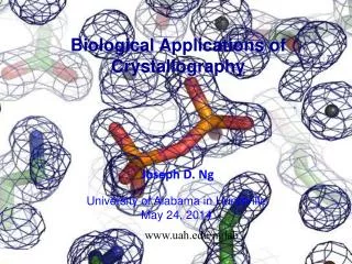
Examination of Protein Crystallography Applications Kathleen Kerr Bragdon, Ph.D. Supervisory Patent Examiner Art Unit 1656
Overview • What is protein crystallography subject matter? • Basis for discussion: Trilateral Co-operation Biotechnology Project • Statutory Subject Matter - 35 USC § 101 • Written Description and Enablement - 35 USC § 112, first paragraph • Prior Art - 35 USC § 102 and 103 2
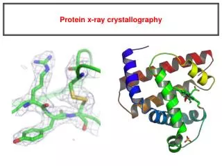
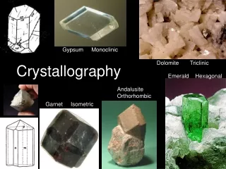
Protein Crystallography Subject Matter • Protein crystallography The art of getting a protein to “sit still” …then taking a 3D “picture” • What are protein crystals? Static, well-ordered arrays of protein molecules • How are these “pictures” made? By projecting x-rays through the ordered protein arrays, collecting the constructively diffracted x-rays, and reconstructing a likely model of the protein’s 3D structure 3
Biotechnology in 3-Dimensions • Useful for identifying inhibitors & agonists 4
From Protein Data Bank (PDB) file 1HSG Crystal Structure at 1.9 A Resolution of HIV II Protease J.Biol.Chem.v269pp.26344-26348 , 1994 5
HEADER HYDROLASE (ACID PROTEINASE) 31-MAR-95 1HSG 1HSG 2COMPND 2 MOLECULE: HIV-1 PROTEASE; 1HSG 4COMPND 3 CHAIN: A, B; 1HSG 5SOURCE 2 ORGANISM_SCIENTIFIC: HUMAN IMMUNODEFICIENCY VIRUS TYPE 1; 1HSG 10SOURCE 3 GENE: HIV-1 PROTEASE FROM THE NY5 ISOLATE; 1HSG 11EXPDTA X-RAY DIFFRACTION 1HSG 13REMARK 2 1HSG 25REMARK 2 RESOLUTION. 2.0 ANGSTROMS. 1HSG 26REMARK 3 R VALUE 0.166 1HSG 31REMARK 3 RMSD BOND DISTANCES 0.017 ANGSTROMS 1HSG 32REMARK 3 RMSD BOND ANGLES 1.9 DEGREES 1HSG 33SEQRES 1 A 99 PRO GLN ILE THR LEU TRP GLN ARG PRO LEU VAL THR ILE 1HSG 62ATOM 1 N PRO A 1 29.361 39.686 5.862 1.00 38.10 1HSG 107ATOM 2 CA PRO A 1 30.307 38.663 5.319 1.00 40.62 1HSG 108ATOM 3 C PRO A 1 29.760 38.071 4.022 1.00 42.64 1HSG 109ATOM 4 O PRO A 1 28.600 38.302 3.676 1.00 43.40 1HSG 110ATOM 5 CB PRO A 1 30.508 37.541 6.342 1.00 37.87 1HSG 111ATOM 6 CG PRO A 1 29.296 37.591 7.162 1.00 38.40 1HSG 112ATOM 7 CD PRO A 1 28.778 39.015 7.019 1.00 38.74 1HSG 113ATOM 8 N GLN A 2 30.607 37.334 3.305 1.00 41.76 1HSG 114ATOM 9 CA GLN A 2 30.158 36.492 2.199 1.00 41.30 1HSG 115ATOM 10 C GLN A 2 30.298 35.041 2.643 1.00 41.38 1HSG 116ATOM 11 O GLN A 2 31.401 34.494 2.763 1.00 43.09 1HSG 117ATOM 12 CB GLN A 2 30.970 36.738 0.926 1.00 40.81 1HSG 118ATOM 13 CG GLN A 2 30.625 35.783 -0.201 1.00 46.61 1HSG 119ATOM 14 CD GLN A 2 31.184 36.217 -1.549 1.00 50.36 1HSG 120ATOM 15 OE1 GLN A 2 32.006 35.518 -2.156 1.00 53.89 1HSG 121ATOM 16 NE2 GLN A 2 30.684 37.339 -2.061 1.00 51.46 1HSG 122ATOM 17 N ILE A 3 29.160 34.436 2.919 1.00 37.80 1HSG 123ATOM 18 CA ILE A 3 29.123 33.098 3.397 1.00 34.13 1HSG 124ATOM 19 C ILE A 3 28.968 32.155 2.198 1.00 33.19 1HSG 125ATOM 20 O ILE A 3 28.088 32.330 1.368 1.00 32.74 1HSG 126 6
HEADER HYDROLASE (ACID PROTEINASE) 31-MAR-95 1HSG 1HSG 2COMPND 2 MOLECULE: HIV-1 PROTEASE; 1HSG 4COMPND 3 CHAIN: A, B; 1HSG 5SOURCE 2 ORGANISM_SCIENTIFIC: HUMAN IMMUNODEFICIENCY VIRUS TYPE 1; 1HSG 10SOURCE 3 GENE: HIV-1 PROTEASE FROM THE NY5 ISOLATE; 1HSG 11EXPDTA X-RAY DIFFRACTION 1HSG 13REMARK 2 1HSG 25REMARK 2 RESOLUTION. 2.0 ANGSTROMS. 1HSG 26REMARK 3 R VALUE 0.166 1HSG 31REMARK 3 RMSD BOND DISTANCES 0.017 ANGSTROMS 1HSG 32REMARK 3 RMSD BOND ANGLES 1.9 DEGREES 1HSG 33SEQRES 1 A 99 PRO GLN ILE THR LEU TRP GLN ARG PRO LEU VAL THR ILE 1HSG 62ATOM 1 N PRO A 1 29.361 39.686 5.862 1.00 38.10 1HSG 107ATOM 2 CA PRO A 1 30.307 38.663 5.319 1.00 40.62 1HSG 108ATOM 3 C PRO A 1 29.760 38.071 4.022 1.00 42.64 1HSG 109ATOM 4 O PRO A 1 28.600 38.302 3.676 1.00 43.40 1HSG 110ATOM 5 CB PRO A 1 30.508 37.541 6.342 1.00 37.87 1HSG 111ATOM 6 CG PRO A 1 29.296 37.591 7.162 1.00 38.40 1HSG 112ATOM 7 CD PRO A 1 28.778 39.015 7.019 1.00 38.74 1HSG 113ATOM 8 N GLN A 2 30.607 37.334 3.305 1.00 41.76 1HSG 114ATOM 9 CA GLN A 2 30.158 36.492 2.199 1.00 41.30 1HSG 115ATOM 10 C GLN A 2 30.298 35.041 2.643 1.00 41.38 1HSG 116ATOM 11 O GLN A 2 31.401 34.494 2.763 1.00 43.09 1HSG 117ATOM 12 CB GLN A 2 30.970 36.738 0.926 1.00 40.81 1HSG 118ATOM 13 CG GLN A 2 30.625 35.783 -0.201 1.00 46.61 1HSG 119ATOM 14 CD GLN A 2 31.184 36.217 -1.549 1.00 50.36 1HSG 120ATOM 15 OE1 GLN A 2 32.006 35.518 -2.156 1.00 53.89 1HSG 121ATOM 16 NE2 GLN A 2 30.684 37.339 -2.061 1.00 51.46 1HSG 122ATOM 17 N ILE A 3 29.160 34.436 2.919 1.00 37.80 1HSG 123ATOM 18 CA ILE A 3 29.123 33.098 3.397 1.00 34.13 1HSG 124ATOM 19 C ILE A 3 28.968 32.155 2.198 1.00 33.19 1HSG 125ATOM 20 O ILE A 3 28.088 32.330 1.368 1.00 32.74 1HSG 126 7
A ball-and-stick model of HIV II protease active site residues complexed with L-735,524 which is an orally bioavailable inhibitor of the HIV protease J.Biol.Chem.v269pp.26344-26348 , 1994 8
Biotechnology in 3-Dimensions • Useful for identifying inhibitors & agonists • Useful for identifying protein-protein and/or protein/DNA interactions 9
From Protein Data Bank (PDB) file 1YCS Structure of the p53 tumor suppressor bound to the ankyrin and SH3 domains of 53BP2. Sciencev274pp.1001-1005 , 1996 10
From Structure of the p53 tumor suppressor bound to the ankyrin and SH3 domains of 53BP2. Sciencev274pp.1001-1005 , 1996 11
From Structure of the p53 tumor suppressor bound to the ankyrin and SH3 domains of 53BP2. Sciencev274pp.1001-1005 , 1996 12
Biotechnology in 3-Dimensions • Useful for identifying inhibitors & agonists • Useful for identifying protein-protein interactions • Useful in proteomics 13
Identifying a fold of the SARS ADRP Domain. A is a bovine Leu-aminopeptidase, B is E. coli pepA, C is a yeast Appr phosphatase, D is E. coli hypothetical protein Er58, E is Archeoglobus fuldiges AF1521, and F is ADRP domain of SARS nsp3. Structure v13, pp.1665-1675, 2005 14
Identifying the active site of the SARS ADRP Domain. yeast Appr phosphatase homolog is in purple, Archeoglobus fuldiges AF1521 in cyan, and SARS nsp3 in green with a ball-and-stick depiction of ADP-ribose in the active site. Structure v13, pp.1665-1675, 2005 15
Biotechnology in 3-Dimensions • Useful for identifying inhibitors & agonists • Useful for identifying protein-protein and/or protein/DNA interactions • Useful in proteomics • Useful in producing “designer” proteins 16
Trilateral Co-operationBiotechnology Project WM4Protein 3-Dimensional (3d) structure related claims • November, 2002 • EPO, JPO, and USPTO input • Addressing “increasing numbers of applications claiming inventions related to … 3-D structural information” 17
35 USC § 101Statutory Classes of Invention 35 USC § 101 reads “Whoever invents or discovers any new and useful process, machine, manufacture, or composition, or any new and useful improvement thereof, may obtain a patent therefore...” (emphasis added) Categories =process machine manufacture composition of matter 18
35 USC § 101Statutory Classes of Invention - Examples Claim 1. A data array comprising the atomic coordinates of protein P as set forth in Figure 1. The 3-D coordinates of a protein constitute nonfunctional descriptive material. Claim 2. A computer model of protein P generated from the data array of Claim 1. Claim 3. A computer-readable storage medium encoded with the data array of Claim 1. Claim 4. A computer comprising the data array of Claim 1 stored in memory. Claim 5. The computer of Claim 4, additionally comprising executable code for: (a) displaying the data array as a 3-dimensional model; (b) analyzing the binding site of the model of protein P; (c) screening in silico a library for small molecules that fit into said binding site; and (d) controlling a unit for assaying the small molecules determined in step (c) in a protein P binding assay. Claims 1-3 paraphrased from Trilateral Project WM4, Cases 1 and 2. See also MPEP 2106 19
35 USC § 101Statutory Classes of Invention - Examples Claim 6. A data array comprising the atomic coordinates of protein P as set forth in Figure 1. Claim 7. An isolated protein P having the structure defined by the structural coordinates of the data array of Claim 6. Claim 8. A pharmacophore having a spatial arrangement of atoms defined by the binding pocket identified in the data array of Claim 6. A pharmacophore is a description of a generalized concept of molecular features in terms of information on spatial arrangement of chemical elements (e.g. hydrophobic groups, ionizable groups, H bond donors/acceptors, etc.) Claim 9. An isolated compound or its salt defined by the pharmacophore of Claim 8. 20 Claims paraphrased from Trilateral Project WM4, Cases 1, 3, and 8
35 USC § 112, first paragraphWritten Description 35 USC § 112, first paragraph reads “The specification shall contain a written descriptionofthe invention, and of the manner and process of making and using it, in such full, clear, concise, and exact terms as to enable any person skilled in the art to which it pertains, or with which it is most nearly connected, to make and use the same and shall set forth the best mode contemplated by the inventor of carrying out his invention.”(emphasis added) 21
35 USC § 112, first paragraphWritten Description - Examples Claim 10. An isolated and purified protein P having the structure defined by the structural coordinates as shown in Figure 1. Figure 1 teaches a “complete” 3D structure of full-length protein P. Claim 11. An isolated and purified protein P having: • a molecular weight of 315 kD as measured by SDS-PAGE, • a pI of 7.5, • an N-terminal amino acid sequence of SEQ ID NO:10, and • the activity of full length protein P. 22 Claim 10 paraphrased from Trilateral Project WM4, Case 3
35 USC § 112, first paragraphWritten Description - Examples Claim 12. An isolated and purified protein P having at least the structure defined by the binding pocket amino acids identified in Figure 2. Figure 2 teaches a partial 3D structure of protein P, limited to the binding pocket amino acids, which are about only 10% of the entire protein. Claim 13. An isolated and purified protein P having: • a protease fragment with a molecular weight of 31 kD as measured by SDS-PAGE, • a protease fragment with a pI of 7.5, • an N-terminal amino acid sequence of SEQ ID NO:10, and • the activity of full length protein P. 23 Claim 12 paraphrased from Trilateral Project WM4, Case 5
35 USC § 112, first paragraphEnablement 35 USC § 112, first paragraph reads “The specification shall contain a written description of the invention, and of the manner and process of making and using it, in such full, clear, concise, and exact terms as to enable any person skilled in the art to which it pertains, or with which it is most nearly connected, to make and use the same and shall set forth the best mode contemplated by the inventor of carrying out his invention.” (emphasis added) 24
35 USC § 112, first paragraphEnablement - Examples Claim 14. The crystalline form of protein P having unit cell dimensions a=4.0 nm, b=7.8nm, and c=11.0nm. The specification teaches the recombinant expression and purification of the claimed protein P as defined by SEQ ID NO:2 (which includes a His-tag for ease of purification in E. coli). This purified protein sample was then subjected to clearly described crystallization conditions to produce x-ray quality crystals of a particular unit cell dimension (e.g. the size of the repeating unit in the ordered array) and space group P2121 (e.g. the organization of the repeating unit). Claim paraphrased from Trilateral Project WM4, Case 4 25
35 USC § 112, first paragraphEnablement - Examples Claim 15. An isolated and purified protein having the sequence shown in SEQ ID NO:1. Claim 16. The protein of Claim 15 in crystalline form. Claim 17. The protein of Claim 15 in soluble form. Claims paraphrased from Trilateral Project WM4, Cases 3 and 4 26
35 USC § 112, first paragraphEnablement - Examples Claim 18. A method of identifying compounds that bind protein P comprising: (a) obtaining a 3-D molecular model of protein P as shown in Figure 1; (b) reducing said model to a 3-D molecular model of the binding pocket of protein P as shown in Figure 2; (c) comparing the model of (b) with a library of 3-D molecular models representing structures of candidate compounds to electronically screen said library; (d) identifying candidate compounds whose structures electronically fit in the model of (b) as compounds that can bind protein P; and (e) assaying the binding of candidate compounds identified in step (d) using purified protein P; to thereby identify compounds that bind protein P. Claim paraphrased from Trilateral Project WM4, a combination of Cases 6 and 7 27
35 USC § 102 and 103Prior Art 35 USC § 102(b) reads “A person shall be entitled to a patent unless – the inventionwas patented or described in a printed publication in this or a foreign country or in public use or on sale in this country, more than one year prior to the date of application for patent in the United States.” (emphasis added) 35 USC § 103(a) reads “A patent may not be obtained though the invention is not identically disclosed or described as set forth in section 102 of this title, if the differences between the subject matter sought to be patented and the prior art are such that the subject matter as a whole would havebeen obviousat the time the invention was made to a person having ordinary skill in the art to which said subject matter pertains.” (emphasis added) 28
35 USC § 102 and 103Prior Art – Examples Claim 19. An isolated and purified protein P having the structure defined by the structural coordinates as shown in Figure 1. Claim 20. The protein of Claim 19 in crystalline form. 29 Claims paraphrased from Trilateral Project WM4, Case 3 and 4
35 USC § 102 and 103Prior Art – Examples Claim 21. A method of identifying compounds that bind protein P comprising: (a) obtaining a model of protein P as shown in Figure 1; (b) using said model in a method of rational drug design to identify candidate compounds that can bind protein P; and (c) assaying the binding of candidate compounds identified in step (b) using purified protein P; to thereby identify compounds that bind protein P. 30 Claim paraphrased from Trilateral Project WM4, a combination of Cases 6 and 7
References • Trilateral Co-operation Biotechnology Project on 3-dimensional proteins http://www.trilateral.net/projects/biotechnology/protein_3d/ • USPTO Guidelines for Computer-related inventions http://www.uspto.gov/web/offices/pac/compexam/comguide.htm • In re Gulack, 217 USPQ 401 (Fed. Cir. 1983) • In re Ngai, 70 USPQ2d 1862 (Fed. Cir. 2004) • In re Lowry, 32 USPQ2d 1031 (Fed. Cir. 1994) • In re Warmerdam, 31 USPQ2d 1754 (Fed. Cir. 1994) • State Street Bank & Trust Co. v. Signature Financial Group Inc., 47 USPQ2d 1596 (Fed. Cir. 1998) • NCBI Structure Database http://www.ncbi.nlm.nih.gov/Structure/ • Protein Data Bank (PDB) http://www.rcsb.org/pdb/home/home.do 31
Acknowledgements Nashaat Nashed Ardin Marschel David Steadman Jean Witz Suzanne Noakes Alexander Kim Jae Wan Lee 32