Download
1 / 37
400 likes | 550 Vues
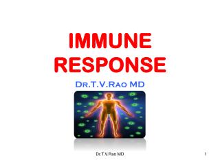
IMMUNE RESPONSE: INFECTION AND VACCINATION. Activation of naïve T cells. Naïve T cells are activated in lymph nodes and spleen. Dendritic cells are key antigen presenting cells for naïve T cells.

E N D
Activation of naïve T cells • Naïve T cells are activated in lymph nodes and spleen. • Dendritic cells are key antigen presenting cells for naïve T cells. • Naïve T cells require MHC/peptide plus antigen-presenting cell "costimulation" in the form of B7 molecules. • Only "professional" antigen presenting cells express B7 molecules. And these only express B7 when activated. • Antigen stimulation without costimulation can lead to loss of T cell responsiveness and immune tolerance.
How naïve, mature abT cells find antigen • Antigen transport. Naïve cells are mainly in lymph nodes and spleen. Dendritic cells transport antigen to draining lymph nodes, blood borne antigens lodge in spleen. • T cell entry to lymph nodes. T cells recirculate through lymph nodes using homing receptors, integrins and responding to chemokines constitutively produced by the lymph node stroma. (Parham 6.2-6.3) • T cell scan of APCs. T cells scan antigen presenting cells. Interaction is initiated by adhesion molecules. (Parham 6.4) • Antigen recognition.
Recognition by T cells of peptide/MHC on dendritic cell leads to stronger adhesion and a more prolonged interaction.
CD8 CD4 CD8 CD4 1 1 inappropriate activation leads to cell inactivation inappropriate activation leads to cell inactivation 2 CD4 Th2 CD8 killer CD4 Th1 Diverse outcomes of immune activation of mature abT cells naive Activation, proliferation, differentiation effectors Th1 vs Th2 is defined by distinctive pattern of cytokine expression How are these cell fate decisions made?
T cells encountering antigen on non-activated professional antigen presenting cells become inactivated inactivated
Figure 6-10 Major professional antigen presenting cells.
Costimulation by signaling through CD28 Appropriate activation of naïve T cells requires in addition to a ligand for the T cell receptor a second interaction with CD28. This second signal is called "costimulation." Under many conditions, dendritic cells provide this signal during a primary response.
Special importance of dendritic cells in activating naïve T cells • It is currently believed that naïve T cells require activation by dendritic cells, macrophages or other "professional" antigen presenting cells. • Dendritic cells are most efficient because they not only take up and present antigen, but also because they move efficiently to lymph nodes to encounter naïve T cells. • These professional antigen presenting cells provide peptide MHC complexes along with a critical "costimulation." • Costimulation is believed to be a crucial interaction between the innate and adaptive immune systems that regulates T cell responses.
Figure 6-2 Immature dendritic cells in tissues actively capture, take up, and process antigens. Green, MHC Upon maturation, they migrate to draining lymph nodes and present antigens to T cells. Red, Lysosomes
Figure 6-12 Activated Activated Dendritic cells express B7 molecules
Only activated antigen presenting cells express B7 molecules which are among the most important costimulatory signals.
In macrophages and dendritic cells, innate immune signals stimulate B7 expression.
Figure 6-15 Inert proteins do not stimulate B7 expression, even if they are foreign.
Adjuvant: important component of vaccines No detectable antibody response Ovalbumin Strong antibody response Ovalbumin + Adjuvant Adjuvants work by slowing the release of antigen, promoting phagocytosis, and providing inflammatory signals that promote costimulation
Figure 12-4 Active ingredient N-actylmuramyl-L-alanine-D-isoglutamine (MDP) *** ** **
Figure 6-15 Inert proteins given with adjuvants can stimulate a response.
Diptheria/ tetanus/ pertussis vaccine Mixture of diptheria toxin (a soluble protein) tetanus toxin (another soluble protein) and killed Bordetella pertussis bacteria. The presence of the bacteria stimulates an improved response to the toxins (presumably by upregulating B7 expression by antigen presenting cells). It also leads to more inflammation and discomfort at the site of injection. Current adjuvant research is trying to identify ways of improving the efficacy of vaccine adjuvants while at the same time reducing unwanted side effects.
Cross presentation for CD8 priming • Problem: how can naïve CD8 cells get activated if activation requires peptide/MHC class I plus costimulation? • This would require that professional antigen presenting cells get infected with virus, for example. • A second problem is that viruses that manage to suppress MHC class I would evade detection. • Cross presentation of exogenous proteins on MHC class I can be specifically carried out by dendritic cells.
Dendritic cell "cross presentation" of ingested antigens on MHC class I There is an important exception to the notion that class I MHC molecules present peptides generated within the cytoplasm. What if the virus doesn’t infect macrophages or dendritic cells? How do viral peptides get presented by professional antigen presenting cells so that an immune response can be induced? The answer is that dendritic cells, but not other cells, have a specialized mechanism for shuttling peptides taken up by endocytosis or phagocytosis into the MHC class I pathway.
CD36 and avb5 Take up apoptotic cells for processing within the dendritic cell
Cross presentation = Dendritic cell, but not other cells, can take up antigens and shuttle them into the class I pathway. This is most efficient when the antigens are particulate Provides a mechanism to get viral and bacterial antigens into profession antigen presenting cells. This induces an immune response to the offending pathogens. Trends in Immunology 22:141-148 (2001)
In some cases, CD8 T cells require T cell help for activation
Failsafe Note that in almost all cases, two (or more) distinct cell types must be independently activated in order for a productive immune response to occur. In all cases, both innate and antigen-specific stimulus is required. 1) Ag+MHC Class II 2) B7 coexpressed by APC 1) Ag+MHC Class I 2) B7 coexpressed by APC 1) Ag+MHC Class I 2) CD4effector Ag+MHC Class II 1) Ag:sIg crosslinking 2)CD4effector Ag+MHC Class II 1) Antigen specific 2) Innate signals 1) Antigen specific 2) Innate signals 1) Antigen specific 2) Antigen specific 1) Antigen specific 2) Antigen specific CD4 CD8 CD8 B
Figure 6-18 IL-2 is an important T cell cytokine
In the absence of appropriate costimulation, T cells can become inactivated (anergic).
B7 Manipulation of costimulation for therapy Tumor immunity Tumors typically express MHC class I molecules, but fail to provoke an immune response because of CD4 or CD8 T cell tolerance. Some tumors do express unique antigens (such as mutated oncogenes) that could be used to direct killer T cells, but they fail to express costimulatory molecules. • Approaches • Force expression of B7 molecules in antigen tumor cells by gene therapy. • Immunize patients with their own tumors in the presence of powerful adjuvants.
Concepts • Naïve T cells encounter antigen in the draining lymph nodes and spleen, not at the site of infection. • Antigen encounter involves transport of antigen, often by uptake of dendritic cells followed by their migration. • Dendritic cells are key antigen presenting cells for naïve T cells. Other important professional antigen presenting cells are macrophages and B cells. • Naïve T cells require peptide/MHC plus costimulation. • A major source of costimulation is B7 expressed by activated professional antigen presenting cells. • B7 expression by dendritic cells and macrophages is often stimulated by innate immune ligands. • Encounter of antigen in the absence of costimulation leads to tolerance. And this is a major pathway of self-tolerance to peripheral antigens. • Crosspresentation of exogenous antigens on MHC class I molecules is carried out by dendritic cells, but no other cells. • Inducing costimulation is an important aspect of vaccine adjuvants.