Download
1 / 55
550 likes | 553 Vues
This lecture covers various biomedical devices such as endoscopes, lasers, electrosurgery devices, and lithotripters. It discusses the types of endoscopes, their illumination and observation methods, and the applications of fiberscopes.

E N D
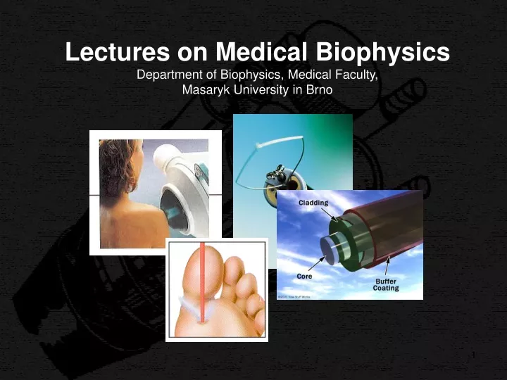
Lectures on Medical BiophysicsDepartment of Biophysics, Medical Faculty, Masaryk University in Brno
Lectures on Medical BiophysicsDepartment of Biophysics, Medical Faculty, Masaryk University in Brno Endoscopes, tissue ablation devices and lithotripters
Lecture content This lecture deals with the following biomedical devices: • Endoscopes • Lasers • Electrosurgery devices • Ultrasonic devices • Cryosurgery devices • Water jet surgery devices • Lithotripters Keep in mind that endoscopes are often equipped with surgical tools including lasers (mainly for tissue ablation). Lithotripsy is a minimally invasive method for removal of kidney stones and gallstones (helps avoid major abdominal surgery).
Endoscopy • Endoscopes are devices for the visual examination of body cavities. They are based on the reflection and refraction of light. • They are inserted into the body cavity to be examined either through natural body openings (nasal and pharyngeal cavity, larynx, airways, urethra, uterus, rectum) or surgical incisions (abdomen, thorax, joints). • Endoscopes can be categorized according to their complexity, method of illumination and method of observation. • There are groups of endoscopes with different complexity: • Endoscopic mirrors • Endoscopes with rigid tubes • Fiberscopes and videoendoscopes • Endoscopic capsules • Endoscopes are used also for minor surgery as they can be equiped with small surgical tools.
Way of illumination and observation • Lighting can be: • Internal: source of light is part of the device • External: examined cavity is illuminated by an external source (Endoscopic mirrors are typical representatives of the second group). • In endoscopes with internal lighting, the source is directly inside the body cavity(distal lighting) or outside the cavity (light is guided into the cavity by an optical system, proximal lighting). • The observation of the body cavity can be: • direct when the physician uses his/her own eyes aided by an optical system • indirect when the images are taken by a digital video camera and observed on a monitor
Endoscopic mirrors (specula) • Laryngoscope. Spoon-like mirror used for the examination of the larynx and posterior part of the nasal cavity. • Otoscope. Funnel-like endoscope inserted into the auditory meatus to examine its distal part and the ear drum. • Rhinoscope. Pliers-like instrument with concave reflecting jaws – examination of anterior part of nasal cavity. • Ophtalmoscopic mirror. Planar or concave mirror with an central orifice. It serves for induction of the so called red reflex – reflection of light from the retina. • Retina is examined by direct ophtalmoscopy – an ophtalmoscope, a small see-through endoscope with light source and correction of the doctor’s visual handicap. • Vaginal speculum (colposcope). Pliers-like instrument with concave reflecting jaws – examination of vagina and cervix.
Endoscopic mirrors Rhino-scope laryngoscope otoscope
Endoscopic mirrors ophtalmoscope vaginal speculum
Rigid tube endoscopes • Rigid metallic tubes with optical system and built-in light source (proximal or distal). Disadvantages: relatively high light loss and the rigidity of tubes. • Cystoscope – urinary bladder • Rectoscope – rectum and sigmoid colon • Endoscopes inserted surgically: • Laparoscope – abdominal cavity. • Arthroscope – joints (namely knee joint).
Rigid tube endoscope rectoscope cystoscope
Fiberscopes • trachea and bronchi (bronchoscopy) • oesophageal mucosa(Oesophagoscopy) • gastric mucosa (Gastroscopy) • colon (colonoscopy) Fibre optics, total reflection, critical angle. The lowest light loss is typical for two-layer optical fibres made of glass or plastics. The core has higher index of refraction n1 than the coating n2. Total reflection occurs when sina < (n12 - n22)1/2. The fibres form bundles serving for illumination and image transfer. In the image transferring bundle, the fibres are arranged in the same way both on input and the output of the bundle. Light signal loss: 0,001 - 0,005 dB per 1 m of length.
Fiberscopes • The fiberscopes make possible to take tissue samples and to make minor surgery. The are flexible so we can examine body parts which are not accessible by rigid endoscopes. Length 130 - 140 cm. • Inside the flexible cable we can see: • 3 bundles of optical fibres (2 for illumination, 1 for image transfer), • a tube for air or water, • a channel for insertion of surgical tools and • control drawbars enabling movement of the distal end with objective giving a sharp image from the distance of 3 - 100 mm. • The proximal end is equipped by an eyepiece mounted in the rigid part of the tube. There is also the control device for distal end movement. • A powerful source of light, air and water pump and vacuum pump are also parts of the device.
Fiberscopes Frontal part of the colonoscope - www.endoscopy.ru/diler/ pentaxvideo.html.
Video-endoscopy Videoendoscopy– modern endoscopes with a video camera. The image is shown on a monitor. http://www.bethesda.de/kliniken/medizinische-klinik-ii---gastroenterologie/endoskopien-spiegelungen/index.php
Laser • Light Amplification by Stimulated Emission of Radiation. • The first ruby laser was constructed by T.H. Maimann in 1960. Main parts of a laser: • active medium • optical resonator • source of excitation energy • Principle of the laser: alternating excitation and deexcitation. • Electrons of the atoms of the active medium are excited (brought to a higher energy level) by the source energy („optical pumping“). • Thereafter they are deexcited by a stimulating photon, new photons of the same energy arise and the effect is repeated – amplification occurs. • In the so-called three-level laser, the third energy level is broad, thus it is not necessary to use monochromatic (i.e. monoenergetic) light for optical pumping. Because of small energy difference between the second and third energy level, the electron transition to the second energy level is spontaneous („thermal“) – electrons are waiting for the stimulating photon there.
Three-level laser Scheme of the 1st ruby laser http://www.llnl.gov/nif/library/aboutlasers/Ruby%20cutaway.GIF
Lasers • Solid l. (compact, semiconductor): ruby laser (694,3 nm), neodymium (1,06 µm), • Semiconductor l.– based on the principle of electroluminescence. • Liquid l. An organic dye solution is used as active medium. Advantage: can be tuned to different wavelengths (from near IR, VISto UV range). • Gaseous l.. Important for medicine. Helium-neon laser (1,06 µm) and ion lasers (argon and krypton). CO2-N2-He-laser etc. • Plasma l. Active medium is plasma, fully ionised carbon – irradiates soft X-rays. • Lasers can operate in two modes:continuousandpulsed • Laser power ranges from 10-3 to 104 W. Low-power lasers (soft-lasers) are used mainly in physical therapy. High-power lasers are used as surgical tools (laser scalpel).
Effects of laser radiation • Laser light is monochromatic and coherent. This allows us to concentrate the laser beam on a small area and to reach a high output density, that makes this surgical instrument useful even in microsurgery. The laser beam can be guided by mirrors, lenses, or optical fibres. Photons are absorbed in the surface layers of tissues. • Thermaleffects depend on the power density of light and its wavelength. They are exploited mainly in surgery and microsurgery. Non-thermaleffects are typical for soft-lasers, they depend little on the wavelength – based on a molecular action mechanism (action on enzymes of the respiratory chain, enhancement of mitochondrial DNA replication, enhancement of enzyme activity). Membrane potentials are also affected, possibly due to changes in membrane permeability for Na+, K+ a Ca++ ions. • Laser light also has a photodynamiceffect – chemical changes of inactive substances irradiated by laser light of certain wavelength can lead to formation of biologically active (cytotoxic) derivatives.
Laser therapy – Safety • In non-invasive phototherapy, powers below 500 mW are used. Classes of lasers used are: • II (power up to 1 mW), • IIIa (power up to 5 mW) • IIIb (power up to 500 mW). • Surgery: Power lasers IV are used • Safety: • Labels placed on lasers must state class, • from IIIb also warning on eye damage by focussed beam • Medical staff as well as the patient must wear goggles absorbing laser light of given wavelength.
Soft-laser therapy • Surface applications – short wavelength, deep applications – long wavelength (near IR). • laser pens are the simplest devices, based on laser diodes, fed by batteries, constant power setting. • Small lasers (pocket) with exchangeable probe, different frequency modes are possible. • Tabletop lasers – user comfort, many functions and applications.
Laser pen Table-top soft-laser
Soft-laser therapy • Analgesic effect: increase of O2 partial pressure, increase of resting potential lowering of its excitability. • Anti-inflammatory effect should be caused by activation of monocytes and macrophages, increased phagocytosis, increased proliferation of lymphocytes. • Biostimulating effect: referred increased synthesis of collagen, better blood supply, faster regeneration of some tissues. • Indications: laryngology, dentistry, orthopaedics and gynaecology. Seldom used as monotherapy. • Opinion of biophysicists: mostly placebo effect, specific action is supported by little research evidence.
? Surgical laser unit
High-power laser application General surgery: A laser can serve as an optical lancet cutting without contact. The blood vessels are coagulated and the cut practically does not bleed.The cutting speed depends on intensity (output density) and on the properties of the tissue.The most frequently used lasers are infrared, namely CO2 laser (10.6 mm) or solid Nd:YAG laser (1.064 mm). Ophthalmology: Besides being the light source of many optical instruments used for examination, the main use is photocoagulation of retina and photoablation of cornea to correct refraction defects. Lasers used for photocoagulation are mostly Nd:YAG with green light 532 nm, adjustable output up to 1.5 W. For corneal refraction defects removal – photoablation - ArF or KrF excimer (excited dimers) lasers are used. They emit UV radiation with 193 nm wavelength. It causes photochemical ablation of the collagen macromolecules in the cornea (every impulse removes 0.1 - 0.5 mm of the tissue). The aim is to change the curvature of the cornea and its refraction, thus improving the patient's vision.
High-power laser application • In dentistry, neodymium and erbium YAG lasers are used. The Nd:YAG laser (1.064 mm) is used in oral surgery and endodontics. The Er:YAG laser (2.940 mm) is used for precise preparation of the tooth enamel and dentine. • Dermatology uses ruby lasers (690 nm) or other laser types including Nd:YAG and alexandrite lasers (adjustable from 720 to 830 nm, well absorbed by skin melanin). The main applications are photocoagulation of varicose veins, wart removal, skin lifting, depilation and tattoo removal.
Laser applications caries removal Face lifting removal of warts
Electrosurgery • These methods use heating effects of high frequency electrical currents. An electrode with a point or a sharp edge can develop a high density of current. • Heat effects are so extensive that water evaporates in the cells, causing their destruction. The high temperature causes coagulation of the tissues and blood, so no bleeding (haemorrhage) occurs. The operating frequency of electrosurgical instruments is about 3 MHz, the output is adjustable up to 500 W. The power differs according to the aim of the surgical intervention (50 W is used in eye and teeth surgery, higher output in breast and abdominal surgery and traumatology). • Electrosurgery devices are equipped with electrodes for electrocoagulation, which close bleeding vessels by coagulation of proteins.
Electrosurgery Electrosurgical unit Point electrode for removal skin defects
Electrosurgery Whipple procedure. Transection of the neck of the pancreas with electrocautery.
Endoscopic electrosurgery Removal of the polypus from intestinal mucosa Removal of a small gastric tumour
Ultrasonic tools • Ultrasound of high intensities (50-1000 W.cm-2) can be used in surgery for selective tissue destruction. • 1. Focused ultrasound with high frequency (1-3 MHz) for selective destruction of soft tissue structures. These systems are in clinical test for breast tumour ablation. • 2. Low frequency ultrasound (50-20 kHz) has been developed for surgical use. Ultrasound produced by piezoelectric or magnetostrictive generators is transmitted to the tissue by special wave-guides, able to enhance the amplitude of ultrasound oscillations up to 10 times. A steel lancet or removable tip is attached to the end of the wave-guide. The removable tip is used also as an aspiration tube, so that the destroyed tissue can be sucked away (aspired).
Ultrasonic tools Aspirator.The acoustic vibrator contracts and expands due to ultrasonic oscillations. The motion of the tip (stroke) is approximately 200 pm. The end of the tip experiences high velocities and accelerations that produce the effect of fragmenting contacted tissues. Cavitational Ultrasonic Surgical Aspirator. This modified probe includes an extended flue and a vibrating tip for laparoscopic surgery.
Ultrasonic tools Low frequency intensive ultrasound source –phacoemulsifier- is an indispensable aid for eye surgeons in the extraction of opaque eye lenses (cataracts). The emulsified lens is immediately sucked away (aspired).
Ultrasonic tools in dentistry • The main application field: tartar removal - scaling. Ultrasonic scalers are fast and efficient. They consist of two main parts: the source of electric oscillations necessary to driving generator of ultrasound, and a handpiece containing ultrasonic transducer, working at a frequency of about 40 kHz. The transducer is linked to variously shaped working tips. Some devices are equipped with water spray (rinsing and cooling). • Ultrasonic scaling mechanisms: • direct effect of ultrasonic oscillations of the working tip on the deposited tartar • ultrasonic cavitation • ultrasonic microstreaming
A schematic diagram of ultrasonic scaler (up with magnetostrictive, down with piezoelectric transducer) Ultrasonic tools in dentistry
Ultrasonic tools in dentistry • A somewhat simpler and cheaper alternative to the ultrasonic scaler is the sonic scaler. The audible sound oscillations are obtained mechanically with the help of an unbalanced air turbine. • The next tools using ultrasonic oscillations are endodontic root-canal devices. Contrary to rotary tooth-canal tools they oscillate longitudinally with frequency of 30 - 50 kHz. They have either the form of a thin steel screw-shaped tip or a slightly conical tip with diamond coating. The main effective mechanism is mechanical abrasion of the root-canal walls enhanced by ultrasonic cavitation.
Cryosurgery • The temperature -25 °C down to -190 °C creates ice crystals inside cells and in intracellular spaces. Cell lysis occurs when the ice thaws. • The advantage is the limitation of tissue destruction to the frozen area sparing nearby healthy tissue. The freezing has an anaesthetic effect so that the cryosurgical intervention causes little pain. The wound practically does not bleed. The frozen tissue sometimes is fixed to the tool, which can be used to extract it (cryoextraction of the eye lens when the cataract is operated). Applications in eye surgery, urology, oncology, gynaecology and plastic surgery. • Cryosurgical devices use liquid nitrogen (-196 °C) or other gases to reach low temperature. The proper cryosurgical tool – cryocauter- has a freezing part on its distant end. The end part of the cryocauter is changeable and has a different shape according to the procedure performed. A digital thermometer displays the temperature.
Cryosurgery Cryosurgical equipment using nitrous oxide (N2O) and carbon dioxide (CO2)
Cryosurgery cryoablation of a prostatic tumour
Water jet dissector as a surgical tool The device comprises a pressure pump, a high-pressure tube and a manipulation part with the thin jet of 0.1 mm diameter on its end. Pressures in the range from 1.5 to 5.0 MPa are usually used. The cut borders are smooth. The jet is a sterile isotonic solution, sometimes with medicaments added to limit bleeding or resist infection. It is said that it gives excellent control of the cut, which is especially significant at brain and parenchyma-tous organs (liver, spleen).
Lithotripsy • In the early 80s, extracorporeal shock-wave lithotripsy (ESWL) was introduced in clinical practice. Destruction of stones (kidney, biliary) by the action of multiple shockwaves – strong impulses of acoustic pressure. The debris is removed from the body via natural efferent ways. It is a minimally invasive method. • A rapid onset of pressure gradient arises on an interface of two media as a result of difference in acoustic impedances. If the pressure force exceeds the mechanical resistance of a stone, its progressive fragmentation occurs. Pressures of about 108 Pa are necessary. Many shock waves (50 to 4000, on average 1000) must be applied (synchronously with heart beats). • Main parts of the lithotripter: source of shock waves, focussing device, coupling medium, accurate device for stone targeting (ultrasonograph or X-ray device).
time Lithotripsytime-course of a shock wave
Lithotripsy production of shock waves and their focussing Ellipsoidal metallic mirrors. Shock waves are produced in one focus and are reflected to the second focus.
Lithotripsy Destruction of a kidney stone http://www.nlm.nih.gov/medlineplus/ency/imagepages/19246.htm

![[PDF] Temperature Monitoring during Catheter, Electrosurgery & Tissue Ablation | L201](https://cdn4.slideserve.com/9063962/t-301-dt.jpg)