Download
1 / 57
570 likes | 578 Vues
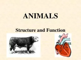
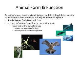
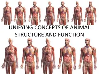
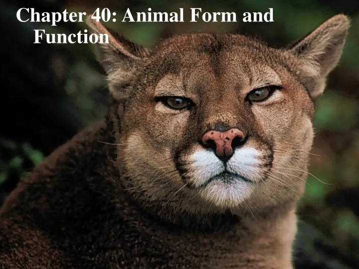
Chapter 40: Animal Form and Function. Animals inhabit almost every part of the biosphere Despite their amazing diversity All animals face a similar set of problems, including how to nourish themselves. Figure 40.1. Form and function are closely correlated.

E N D
Animals inhabit almost every part of the biosphere • Despite their amazing diversity • All animals face a similar set of problems, including how to nourish themselves
Figure 40.1 Form and function are closely correlated
Natural selections select for what works best among the available variations in a population
Evolutionary convergence • Independent adaptation to a similar environmental challenge (a) Tuna (b) Shark (c) Penguin (d) Dolphin Figure 40.2a–e (e) Seal
Exchange with the Environment • Occurs as substances dissolved in the aqueous medium transported across membranes
Diffusion (a) Single cell • Single-celled protist has a sufficient surface area of plasma membrane to service its entire volume of cytoplasm Figure 40.3a
Multicellular organisms with body walls that are only two cells thick facilitate diffusion Mouth Gastrovascular cavity Diffusion Diffusion Figure 40.3b (b) Two cell layers
Organisms with complex body plans • highly folded internal surfaces (lg. surface area) specialized for exchanging materials
External environment Food CO2 O2 Mouth Animal body Respiratory system Blood 50 µm 0.5 cm A microscopic view of the lung reveals that it is much more spongelike than balloonlike. This construction provides an expansive wet surface for gas exchange with the environment (SEM). Cells Heart Nutrients Circulatory system 10 µm Interstitial fluid Digestive system Excretory system The lining of the small intestine, a diges- tive organ, is elaborated with fingerlike projections that expand the surface area for nutrient absorption (cross-section, SEM). Inside a kidney is a mass of microscopic tubules that exhange chemicals with blood flowing through a web of tiny vessels called capillaries (SEM). Anus Unabsorbed matter (feces) Metabolic waste products (urine) Figure 40.4
Animal form and function are correlated at all levels of organization • cells • tissues • organs • organ systems
Tissue Structure and Function • 4 main categories • Epithelial, connective, muscle, and nervous
Epithelial Tissue • Covers the outside of the body and lines organs and cavities within the body • cells are closely joined
Epithelial tissue A simple columnar epithelium A stratified columnar epithelium A pseudostratified ciliated columnar epithelium Stratified squamous epithelia Cuboidal epithelia Simple squamous epithelia Basement membrane Figure 40.5 40 µm
Connective Tissue • Bind and supports other tissues • sparsely packed cells scattered throughout an extracellularmatrix
CONNECTIVE TISSUE • Connective tissue 100 µm Chondrocytes Collagenous fiber Chondroitin sulfate Elastic fiber 100 µm Cartilage Loose connective tissue Adipose tissue Fibrous connective tissue Fat droplets Nuclei 150 µm 30 µm Blood Bone Central canal Red blood cells White blood cell Osteon Plasma Figure 40.5 700 µm 55 µm
Muscle Tissue • Composed of long cells called muscle fibers, contract in response to nerve signals • 3 types: skeletal, cardiac, and smooth
Nervous Tissue • Senses stimuli and transmits signals throughout the animal
Muscle and nervous tissue MUSCLE TISSUE 100 µm Skeletal muscle Multiple nuclei Muscle fiber Sarcomere Cardiac muscle 50 µm Nucleus Intercalated disk Smooth muscle Nucleus Muscle fibers 25 µm NERVOUS TISSUE Process Neurons Cell body Nucleus Figure 40.5 50 µm
Dendrites Cell body Nucleus Synapse Signal direction Axon Axon hillock Presynaptic cell Postsynaptic cell Myelin sheath Synapticterminals Figure 48.5 Neurons
Lumen of stomach Mucosa. The mucosa is an epithelial layer that lines the lumen. Submucosa. The submucosa is a matrix of connective tissue that contains blood vessels and nerves. Muscularis. The muscularis consistsmainly of smooth muscle tissue. Serosa. External to the muscularis is the serosa,a thin layer of connective and epithelial tissue. 0.2 mm • In some organs tissues are arranged in layers Figure 40.6
Organ systems in mammals Table 40.1
Organisms require chemical energy for • Growth, repair, physiological processes, regulation, and reproduction
Bioenergetics • Flow of energy through an animal • Limits the animal’s behavior, growth, and reproduction, how much food it needs
Energy Sources and Allocation • Chemical energy from food food digested molecules generate ATP powers cellular work
Metabolic needs and biosynthesis Organic molecules in food External environment Animal body Digestion and absorption Heat Energy lost in feces Nutrient molecules in body cells Energy lost in urine Cellular respiration Carbon skeletons Heat ATP Biosynthesis: growth, storage, and reproduction Cellular work Heat Figure 40.7 Heat
(a) This photograph shows a ghost crab in arespirometer. Temperature is held constant in thechamber, with air of known O2 concentration flow-ing through. The crab’s metabolic rate is calculatedfrom the difference between the amount of O2entering and the amount of O2 leaving therespirometer. This crab is on a treadmill, runningat a constant speed as measurements are made. (b) Similarly, the metabolic rate of a manfitted with a breathing apparatus isbeing monitored while he works outon a stationary bike. Figure 40.8a, b • Measuring metabolic rate by amount of oxygen consumed or carbon dioxide produced
Birds and mammals are endothermic • bodies warmed by heat generated by metabolism • high metabolic rates
Amphibians, reptiles other than birds, and ………..Daphnia are ectothermic • gain their heat from external sources • lower metabolic rates • Q 10
Size and Metabolic Rate • Metabolic rate inversely related to body size among similar animals
Activity and Metabolic Rate • Basal metabolic rate (BMR) • Metabolic rate of an endotherm at rest • Standard metabolic rate (SMR) • Metabolic rate of an ectotherm at rest
500 A = 60-kg alligator A H 100 H A H = 60-kg human 50 H 10 Maximum metabolic rate (kcal/min; log scale) H H 5 A 1 A A 0.5 0.1 1 minute 1 second 1 hour 1 day 1 week Time interval Key Existing intracellular ATP ATP from glycolysis ATP from aerobic respiration • Animal’s maximum possible metabolic rateis inversely related to the duration of the activity Figure 40.9
Endotherms Ectotherm Reproduction 800,000 Temperature regulation costs Basal metabolic rate Growth 340,000 Activity costs Annual energy expenditure (kcal/yr) 8,000 4,000 4-kg male Adélie penguin from Antarctica (brooding) 60-kg female human from temperate climate 0.025-kg female deer mouse from temperate North America 4-kg female python from Australia (a) Total annual energy expenditures 438 Energy expenditure per unit mass (kcal/kg•day) Human 233 Python Deer mouse Adélie penguin 36.5 5.5 Energy expenditures per unit mass (kcal/kg•day) (b) Energy use Figure 40.10a, b
Animals regulate their internal environment within relatively narrow limits • Homeostasis: balance between external changes and the animal’s internal control mechanisms that oppose the changes
Regulator • Uses internal control mechanisms to moderate internal change in the face of external, environmental fluctuation • Conformer • Allows its internal condition to vary with certain external changes
Response No heat produced Heater turned off Room temperature decreases Set point Too hot Set point Too cold Set point Control center: thermostat Room temperature increases Heater turned on Response Heat produced Homeostatic control system • 3 functional components • receptor, control center, and effector Figure 40.11
Homeostatic control systems function by negativefeedback • buildup of the end product shuts the system off
Positive feedback • change in some variable that amplify the change
Thermoregulation • animals maintain an internal temperature within a tolerable range
40 River otter (endotherm) 30 Body temperature (°C) 20 Largemouth bass (ectotherm) 10 0 10 20 30 40 Ambient (environmental) temperature (°C) Ectotherms • Tolerate greater variation in internal temperature than endotherms Figure 40.12
Endothermy • Energetically more expensive than ectothermy • Buffers animals’ internal temperatures against external fluctuations • High level of aerobic metabolism
Radiation is the emission of electromagnetic waves by all objects warmer than absolute zero. Radiation can transfer heat between objects that are not in direct contact, as when a lizard absorbs heat radiating from the sun. Evaporationis the removal of heat from the surface of a liquid that is losing some of its molecules as gas. Evaporation of water from a lizard’s moist surfaces that are exposed to the environment has a strong cooling effect. Conduction is the direct transfer of thermal motion (heat) between molecules of objects in direct contact with each other, as when a lizard sits on a hot rock. Convectionis the transfer of heat by the movement of air or liquid past a surface, as when a breeze contributes to heat loss from a lizard’s dry skin, or blood moves heat from the body core to the extremities. Heat Exchange • Organisms exchange heat by four physical processes Figure 40.13
Insulation • Thermoregulatory adaptation in mammals and birds • Reduces the flow of heat between an animal and its environment • e.g. feathers, fur, or blubber
Mammal integumentary system • Acts as insulating material Hair Epidermis Sweat pore Muscle Dermis Nerve Sweat gland Hypodermis Adipose tissue Blood vessels Oil gland Figure 40.14 Hair follicle
Circulatory adaptations • Vasodilation • Blood flow in the skin increases, facilitating heat loss • Vasoconstriction • Blood flow in the skin decreases, lowering heat loss
Pacific bottlenose dolphin 1 Arteries carrying warm blood down the legs of a goose or the flippers of a dolphin are in close contact with veins conveying cool blood in the opposite direction, back toward the trunk of the body. This arrangement facilitates heat transfer from arteries to veins (black arrows) along the entire length of the blood vessels. Canada goose Blood flow 1 Artery Vein Vein 2 Near the end of the leg or flipper, where arterial blood has been cooled to far below the animal’s core temperature, the artery can still transfer heat to the even colder blood of an adjacent vein. The venous blood continues to absorb heat as it passes warmer and warmer arterial blood traveling in the opposite direction. Artery 3 3 35°C 33° 30º 27º 20º 18º 2 10º 9º In the flippers of a dolphin, each artery is surrounded by several veins in a countercurrent arrangement, allowing efficient heat exchange between arterial and venous blood. 2 3 As the venous blood approaches the center of the body, it is almost as warm as the body core, minimizing the heat lost as a result of supplying blood to body parts immersed in cold water. • Countercurrent heat exchangers reduce heat loss 3 1 Figure 40.15
21º 23º 25º 27º 29º 31º Body cavity Skin Artery Vein Blood vessels in gills Capillary network within muscle Heart Artery and vein under the skin Dorsal aorta • Some bony fishes and sharks also possess countercurrent heat exchangers (a) Bluefin tuna. Unlike most fishes, the bluefin tuna maintains temperatures in its main swimming muscles that are much higher than the surrounding water (colors indicate swimming muscles cut in transverse section). These temperatures were recorded for a tuna in 19°C water. (b) Great white shark. Like the bluefin tuna, the great white shark has a countercurrent heat exchanger in its swimming muscles that reduces the loss of metabolic heat. All bony fishes and sharks lose heat to the surrounding water when their blood passes through the gills. However, endothermic sharks have a small dorsal aorta, and as a result, relatively little cold blood from the gills goes directly to the core of the body. Instead, most of the blood leaving the gills is conveyed via large arteries just under the skin, keeping cool blood away from the body core. As shown in the enlargement, small arteries carrying cool blood inward from the large arteries under the skin are paralleled by small veins carrying warm blood outward from the inner body. This countercurrent flow retains heat in the muscles. Figure 40.16a, b
Endothermic insects • countercurrent heat exchangers maintain a high temperature in the thorax Figure 40.17
Cooling by Evaporative Heat Loss • Lose heat through the evaporation of water in sweat • Panting cools bodies
Bathing cools animal Figure 40.18