Download

1 / 44
440 likes | 699 Vues
Bacillus Anthracis. DR.R.S.GOPIKA DEPT OF PATHOLOGY AND MICROBIOLOGY SKHMC. Clinically important Gram positive bacilli. Bacillus Clostridium Corynebacterium. Spore forming Bacillus Clostridium Non spore forming Corynebacterium. BACILLUS. Bacillus anthracis Bacillus cereus

E N D
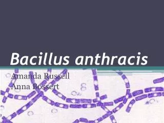
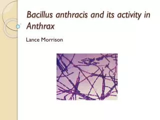
Bacillus Anthracis DR.R.S.GOPIKA DEPT OF PATHOLOGY AND MICROBIOLOGY SKHMC
Clinically important Gram positive bacilli • Bacillus • Clostridium • Corynebacterium
Spore forming Bacillus Clostridium Non spore forming Corynebacterium
BACILLUS Bacillus anthracis Bacillus cereus Bacillus subtilis
Characteristics Gram +ve Rod Aerobic bacilli forming heat resistant spores Peritrichate flagella , motile- except B anthrax Facultative intracellular
Bacillus anthracis Robert Koch-1876 isolated 4-8x1-1.5 m, Single or paired , Encapsulated, bamboo stick -arrangement Non-motile , Spores are oval and central . Spores are formed in culture, dead animal's tissue not in the blood of infected animals.
Culture 12 -45ºC , pH- 7-7.4 Sporulation -25- 30ºC On nutrient agar Grey,granular, 2-3 mm, irregular finger like edge.
Blood agar Colonies have irregular borders and are non-hemolytic. Frosted glass appearance
Blood agar Incubated overnight at 35ºC without carbon dioxide Non-hemolytic, non-pigmented, dry ground glass surface, edge irregular with comma projections, “Medusa Head”. Microscopic-"Medusa head" or "Comet tail".
Gelatin stab agar Inverted fir tree Liquifaction of gelatin more on surface
PLET MEDIA Polymyxin,lysozyme,EDTA,thallous acetate selective media- small smooth colonies Mucoid colonies
M’Fadyeans reaction To identify the capsular materials blood films containing bacteria – stained with poly chrome methylene blue Amorphous purplish material is noticed around the bacteria.
Resistance Vegetative – moist heat at 60ºC – 30 min Dry heat spores- 150ºC -60min Wool- duckering 2% formaldehyde at 30 -40ºC – 20 min Animal hair – 0.25% formaldehyde at 60ºC – 6hrs
Virulence The Capsular Polypeptide Interferes with phagocytosis. Polysaccharide Somatic Antigen Antiboby production – not effective Complex Protein Toxin toxin is responsible for signs and symptoms characteristic of anthrax. Accumulation of the toxin in tissue and its effect on the central nervous system results in death by respiratory failure and anoxia.
TOXINS PA –protective Ag factor – binds on receptors on target cells, helps in entering the cells. OF/EF- oedema factor – adenylatecyclase- activation leading to intracellular accu of cAMP – oedema LF- lethal factor – leads to death of cell –cleaving host cell MAPK (mitogen activated protein kinase )
B. anthracis diseases MoT- Inhalation - Inoculation - Ingestion meat
Three forms: Cutaneous- Most common form acquired through a cut or abrasion of the skin, which comes into contact with spores from the soil or a contaminated animal Inhalation- acquired by the inhalation of spore-containing dust where animal hair or hides are handled Intestinal- consumption of contaminated meat
Cutaneous Anthrax hide porters disease Enters thru cut or abrasion. Cutaneous-Spores germinate, vegetative cells multiply and a lesion, a painless ulcer (1-3 cm) with black necrotic center- Malignant pustule develops at the site of infection
Pathogenesis of Cutaneous Anthrax • 1 to 5 days after contact • Small, pruritic, non-painful papule at inoculation site • Papule develops into hemorrhagic vesicle & ruptures • Slow-healing painless ulcer covered with black eschar surrounded by edema • Infection may spread to lymphatics w/ local adenopathy • Septicemia may develop
Gastrointestinal Anthrax MoT: Digesting undercooked meat containing spores Spores are consumed after eating undercooked meat in the digestive tract germinate and produce bacteria release exotoxins degrading intestinal walls bacteria to spread directly into the bloodstream
25% to 60% mortality rate-100% fatal Clinical Presentation Abdominal pain Inflammation of the intestinal tract, nausea, loss of appetite, vomiting, severe diarrhea Hemorrhagic ascites bacilli gainsentry into the circulation - lead to a life threatening toxemia Paracentesis fluid may reveal gram+ve rods
Inhalation/pulmonary Anthrax Wool sorters disease Inhalation of spores Early presentation mimics an influenza-like illness, with fever, malaise, and cough, progress to fulminant disease and ultimate respiratory collapse,
Inhalation/pulmonary Anthrax spores are inhaled lodge in the alveolar spaces Alveolar macrophages engulf the spores. Spores germinate within macrophages Bacteria proceed to lymph nodes spread into bloodstream begin to release the exotoxins
Symptoms of Inhalation Anthrax Initial symptoms: sore throat, mild fever, muscle aches Severe difficulty breathing Septic shock Development of meningitis Respiratory failure resulting in death
Lab diagnosis Samples are collected depending on the site affected: 1. Swab samples from cutaneous lesions and blood cultures. 2. Sputum and blood for pulmonary anthrax 3. Gastric aspirate, feces and blood for enteric anthrax. Gram stain Culture Serological – Ag demostration
ANTHRACOID BACILLI/PSEUDO ANTHRAX Non pathogenic aerobic spore bearing bacilli resembling B.anthrax Bacillus cereus- pathogenic Bacillus subtilis B.coagulans B.steatothermophilus B.licheniformis B.thuringiensis
BACILLUS CEREUS 1 x 3-4 µm, rod shaped Large, motile, saprophytic bacillus Heat resistant spores cause foodborne illness, "Fried Rice Syndrome"
Cultural characteristics SHEEP BLOOD AGAR. Large colonies are surroundend by a wide zone of beta-hemolysis hemolysis
Bicarbonate agar (left) and blood agar (right) plate cultures of Bacillus cereus
BACILLUS CEREUS AGAR medium used for the detection and enumeration of spores and vegetative forms of Bacillus cereus in food products 5mm in diameter and have a distinctive peacock blue colour surrounded by a egg yolk precipitate of the same colour.
Virulence and Pathogenicity: ability to form endospores Cereolysin- haemolysin phospholipase C group of enzymes – acts on phospholipase C & sphingomyelin of cell membranes. Toxins - enterotoxins and emetic toxins.
Toxins Enterotoxin - 38000-46000 daltons – mucosal damage, fluid accumulation. Heat labile Emetic toxin/cereulide– Pre formed heat and acid stable , effects the potassium ion channel that alters the cell membrane permeability of nerve cells or activate serotonin receptors, leading to increased afferent vagus nerve stimulation
Reproduction At 30 °C (86 °F), a population of B. cereus can double in as little as 20 minutes or as long as 3 hours, depending on the food product.
Food poisoning B. cereus endospores are able to withstand relatively high temperatures, broad pH ranges, and a salt concentration of up to a 10%. The vegetative form starts growing when cooked food is exposed to warm temperatures over a prolonged period of time. The threshold number of B. cereus is around 106 colony forming units (CFU) per gram of food ingested
Pathogenesis Cooking temperatures less than or equal to 100 °C allows some spores to survive. Or when food is then improperly refrigerated, allowing the endospores to germinate. Germination and growth generally occurs between 10–50 °C Bacterial growth results in production of enterotoxins, ingestion leads to two types of illness, diarrheal and emetic syndrome.
Food poisoning Gastroenteritis- diarrhoeal food borne infection heat labile - meat and vegetables causes watery diarrhea, abdominal cramps, and pain. The symptoms can begin 6 to 15 hours after eating contaminated food. Last for 20-36 hrs Emetic form. intoxication - heat stable - Rice causes nausea and vomiting that begins ½ hour to 6 hours after eating the contaminated food. last for 8-10 hours.
Other manifestations Chronic skin infections Ocular infection – post traumatic ophthalmitis Pneumonia Meningitis .
Ospe 1 A 5 year boy was brought to outpatient department complaining of fever and sore throat. On examination his temp. was 38.5°C, the tonsil area and pharynx were obviously inflammed with some foci of pus. What is the differential diagnosis? What investigation should be done?
Osce -2 A 28 Year Old Female after an accident ,presented with a sudden onset of fever, right sided chest pain and productive cough of purulent sputum. On examination her temperature was 39 °C. There were Rhonci and dullness on the right side of the chest X-ray showed massive consolidation on the right side of the chest. What investigation should be done? What is the differential diagnosis?
Osce -3 A 5 year-old boy attended to the OP complaining of sore throat , fever (38.5°C), and a noticed pharyngeal pseudomembrane What is the differential diagnosis? What investigation should be done ?
commons.wikimedia.org en.wikipedia.org Text Book of Microbiology-Anandanarayanan