Download

1 / 32
350 likes | 638 Vues
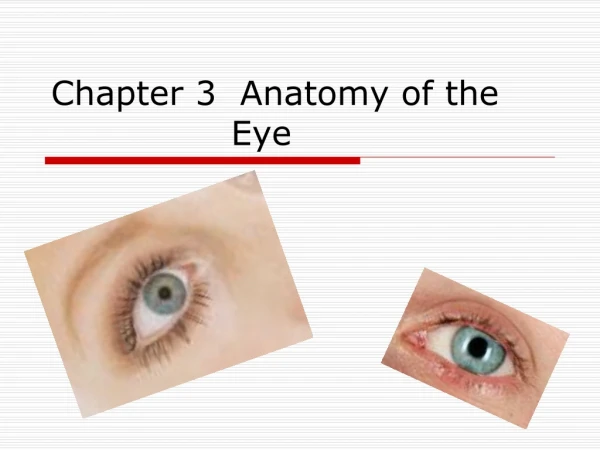
Anatomy of The Eye. The Eye is the organ of vision. Composed of : Eyeball. The adnexa. INTRODUCTION. THE POSITION. In the Predatory species: have set well forward In Herbivores , Ruminant and rabbits: have eyes more laterally to have wide area of vision. Terminology of the eye.

E N D
The Eye is the organ of vision. Composed of : Eyeball. The adnexa. INTRODUCTION
THE POSITION • In the Predatory species: have set well forward • In Herbivores , Ruminant and rabbits: have eyes more laterally to have wide area of vision
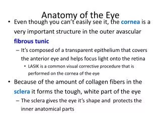
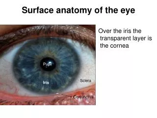
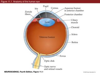
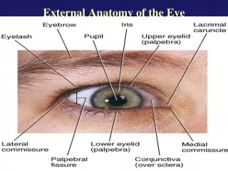
Terminology of the eye • Cornea : the transparent part of the eyeball . • Anterior pole: the highest point on cornea . • Posterior pole : the highest point on posterior surface . • Optic axis: the straight line passing through both poles
The Eyeball • Equator :an imaginary line about the eyeball, which is the equidistant from the poles. • Meridian: is one of many lines passing from pole to pole that intersects the equator at right angles. • Optic nerve :leaves the eyeball slightly ventral to the posterior pole
Eyeball The three tunics are: I- An external fibrous tunic II- A middle vascular tunic III- An internal tunic
Eyeball The three tunics are: I. An external fibrous tunic: that gives form to and protects the eyeball; it’s the only complete tunic. • A middle vascular tunic: that consist largely of blood vessels and smooth muscle • concerned with the nutrition of the eyeball and the regulation of the shape of the lens and size of pupil.
Eyeball III. An internal tunic: that consists largely of nervous tissue • concerned with vision and translation of visual stimuli into nerve impulses for interpretation by the brain.
The Fibrous Tunic • It consists of the sclera and the cornea, which meet at the limbus. 1. The sclera is the opaque posterior part of the fibrous tunic and consists of a dense felt work of colagenous and elastic fibers and is generally white but in some species it contain pigment cells
The fibrous tunic • The cornea forms about one quarter of the fibrous tunic and bulges forward. It is composed off dense connective tissue arranged in lamellar form . • The cornea doesn’t contain blood vessels; nutrients for its cells permeate from vessels in the limbus or are carried to it its surface in the lacrimal fluid and aqueous humor .
The vascular Tunic (uvea) • Deep to the sclera, which it composed of three zones . 1) The choroids: lies on the sclera from the optic nerve to the limbus and contains a dense network of blood vessels embedded in heavily pigmented connective tissue
The vascular Tunic (uvea) • In the dorsal part of the fundus the choroids forms colored, light-reflecting area known as tapetumlucidum • is avascular layer (cellular in the carnivores, fibrous in ruminants and horses) between the capillaries and the vessels. • The tapetum makes the eyes of animals shine when they look toward the light. • Our eyes and those of the pig don’t have a tapetum so they don’t reflect the light. • This reflecting of light is a night vision adaptation because of stimulation of the light sensitive receptors in the retina.
The vascular Tunic (uvea) 2) The ciliary body : toward the limbus the choroids thickness to form it. 3) The Iris: the smallest part of the vascular tunic, which extends from the cornea to the lens. • It attached to sclera and ciliary body by pectinate ligament. • the opening in the center is the pulpi
The vascular Tunic (uvea) • The iris divided the space between the lens and cornea into anterior and posterior chambers tat communicate through pupil and filled with, aqueous humor (a clear watery fluid). • The color of the iris determines the color of the eye • depends on the number of the pigmented cells present in its stroma • the type of the pigment in the cells.
The internal tunic • The internal tunic of the eyeball contains the light-sensitive receptor cells (known as retina). • It’s an extension of the brain to which remains connected by the optic nerve.
The internal tunic The layers in retina are: • A single layer of pigmented cells. • Aneuroepithelialm layer containing the receptor cells, rods and cones and their nuclei. • the rods for black and whit • the cones for the color vision. • A layer of bipolar ganglion cells. • A layer of multipolar ganglion cells nonmyelinated axons lying internal to the cells and pass to the optic disc where they form the optic nerve. • The optic disc is a blind area because there is no receptor cell.
v The adnexa of the eye • The orbital fasciae: a. The periorbital: is attached near the optic foramen at the apex of the cone . b. The superficial muscular fascia: lies within the periorbital. It’s loose and fatty. And envelops in the levatorpalpebraesuperioris and the lacrimal gland. c. The deep muscular fascia: is more fibrous and arises from the eyelids and from the limbus of the eyeball.
v The adnexa of the eye 2. The muscles of the eyeball: • The rectus muscles: dorsal, ventral, medial and lateral are inserted anterior to the equator by wide but very thin tendons. • The ventral and dorsal oblique muscles: attach to the eyeball near the equator.
v The adnexa of the eye 2. The muscles of the eyeball: • The retractor bulbi arises from the vicinity of the eyeball and inserted on the eyeball posterior to the equator. • The levatorpalpebraesuperioris: striated muscle within the orbit that doesn’t attach to the eyeball but passes over it to enter and elevate the upper eyelid
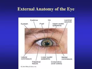
v The adnexa of the eye 3. The eyelids and conjunctiva : • The eyelids (palpebrae) are two musculofibrous folds of which the upper is the more extensive and more mobile. • The free margins of the lids are meet at the medial and lateral angles of the eye and bound an opening known as the palpebral fissure.
v The adnexa of the eye 3. The eyelids and conjunctiva : They are consist of three layers: 1.The skin: is thin and delicate and is covered with short hairs: it may also carry a few prominent tactile airs. 2.The musculofibrous layer: is formed by the orbicularisoculi, the orbital septum, the aponeurosis of the levator muscle and the smooth tarsal muscle. 3.The mucous (palpebral conjunctiva) a thin, transparent mucous membrane
v The adnexa of the eye 3. The eyelids and conjunctiva : • The third eyelid is supported by a T-shaped piece of cartilage. • Bar lies in the free edge of the fold and stem points backward into the orbit medial to the eyeball. • The stem of cartilage is surrounded by lacrimal gland (the gland of the third eyelid).
v The adnexa of the eye • The lacrimal apparatus: • This consists of lacrimal gland proper • The lacrimal gland is flat and lies between the eyeball and the dorsolateral wall of orbit. • The glands associated with the third eyelids • several small accessory glands • duct system that conveys the lacrimal fluid after it has washed over the eye into the nasal cavity for evaporation.
v The blood supply of the eye: • The arteries can be divided into three groups: • THOSE SUPPLY EYEBLL • SUPPLY OCULR MUSCLES • THOSE LAEVING THE ORBIT TO SUPPLY ADJCENT STRCTURES. • The external ophthalmic artery carries the principle supply of the blood to the eye, which is a branch of the maxillary artery.
v The blood supply of the eye: • 1) The branches of the external ophthalmic for the eyeball penetrate the sclera to reach the vascular tunic and retina. • -Short posterior ciliary a. / supply the adjacent choroids in addition to branches to the optic nerve. • -Long posteriorciliary a. /pass close the sclera closer to the equator. • -The anterior ciliary a. / supply the anterior potion of the choroids, the ciliary body and the iris • These arteries anastomose to form the greater arterial circle of the iris.
v The blood supply of the eye: 2) The arteries that supply the ocular muscles. Which the absence of the large vessels in distal ends reduces bleeding when the muscles are cut during the enucleating.
v The blood supply of the eye: 3) The arteries that leave the orbit: • -The lacrimal a. / supply the lacrimal gland in route. • -The supraorbital a. / send branches to the upper eyelids • -The malar a. /supply the eyelids and also adjacent area of the face. • -The external ethamoid a. / supply the ethamoid labyrinth of the nasal cavity.
v The nerve supply of the eye: • The optic nerve II: enters the orbit through the optic foramen and passes to the light receptor cells in the retina. • It allows the movements of the eye and is covered by meninges that it acquired during its development.
v The nerve supply of the eye: • The Oculomoter nerve III: control the movement of the eyeball. it enters the orbit through the orbital fissure. • Supply: dorsal, medial, ventral Rectus muscle • Ventral oblique muscle • Part of retractor muscle • The abducent nerve VI: enters through the orbital foramen and innervates most of retractor bulbi and lateral rectus muscles.
v The nerve supply of the eye: • The trochlear nerve IV: innervate • Dorsal oblique muscle • The trigeminal nerve V: send branches to the eye. • Opthalmic division Give sensory branches to: 1- long ciliary nerve of the eye, lacrimal and supraorbital nerves. • Maxillary division • Zygomatic branch supply ventrolateral segment of the eyelids and conjunctiva
v The nerve supply of the eye: • The facial nerve VII: • passes between the eye and the ear gives auriculopalpebral branch • innervates the orbicularisoculi