Download
1 / 43
430 likes | 459 Vues
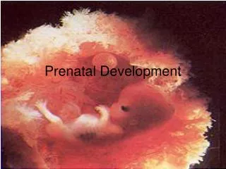
Prenatal development after the eighth week is the fetal period. This is the time when structures grow and specialize. From the start of the ninth week until birth, the prenatal human organism is a fetus. 3.4 Prenatal Development.

E N D
Prenatal development after the eighth week is the fetal period. • This is the time when structures grow and specialize. • From the start of the ninth week until birth, the prenatal human organism is a fetus. 3.4 Prenatal Development A prenatal human is considered an embryo for the first eight weeks. During this time, rudiments of all body parts form. The embryo in the fist week is considered to be in a “preimplantation” stage because it has not yet settled into the uterine lining.
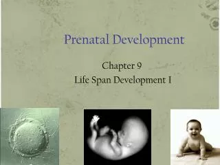
Human Reproduction and Development Three Trimesters of Development • Human development takes around 266 days from fertilization to birth. • The time span is divided into three trimesters.
Human Reproduction and Development The First Trimester • All tissues, organs, and organ systems begin to develop. • At the end of eight weeks, the embryo is called a fetus.
Human Reproduction and Development The Second Trimester • Period of growth • The fetal heartbeat might be heard. The Third Trimester • The fetus continues to grow at a rapid rate. • Fat accumulates under the skin to provide insulation for the fetus once it is born.
Fertilization • Formation of diploid zygote – single cell from fusion of haploid gametes. • Gametes formed by cell division known as meiosis which is a process by which the chromosome content is reduced to 23. (Spermatogenesis or Oogenesis) • Process allows for mixing of maternal and paternal genes Acknowledgement for pictures is courtesy of Alpha (Scientist in Reproductive Medicine Permission confirmed by D Cole 24.5.05).
Human Reproduction and Development Fertilization • Process of a sperm joining with an egg • Sperm and eggs each are haploid, and each normally has 23 chromosomes. • Fertilization restores the diploid number of 46 chromosomes.
Human Reproduction and Development Human Development Before Birth • The tip of each sperm cell is a specialized lysosome called an acrosome that weakens the plasma membrane surrounding the egg. • Eventually the plasma membrane becomes weak enough that one sperm can penetrate the egg. • Immediately following this penetration, the egg forms a barrier to prevent other sperm from entering the now-fertilized egg.
Human Reproduction and Development Early Development • The fertilized egg is called a zygote. • Around 30 hours after fertilization, the zygote undergoes its first mitosis and cell division • By the third day, the embryo, called a morula, leaves the oviduct and enters the uterus.
Mitosis • Mitotic cell division – formation of identical cells • Cells have identical genetic material • Totipotent – clones • Day 4: embryo 16-20 cells & is known as a Morula Acknowledgement for pictures is courtesy of Alpha (Scientist in Reproductive Medicine Permission confirmed by D Cole 24.5.05).
Human Reproduction and Development Human Development Before Birth • By the fifth day, the morula has developed into a blastocyst. • The blastocyst attaches to the endometrium around the sixth day and is fully implanted by Day 10.
Blastocyst • Cells division becomes asymmetrical • Cells polarize • Lose their totipotency and begin to differentiate • Outer cell mass becomes the trophoblast which develops into the placenta • Implants day 6
By day 14 • From implantation of the blastocyst ,the inner cell mass is known as the embryo • Amniotic & Chorionic cavities
Critical time for normal development Particularly sensitive to external factors, environmental hazards, pharmacological agents, drug misuse Organogenesis Trilaminar disc folds into C shaped cylindrical embryo Co-ordinated by genes – Homeobox Cell differentiation Tissue interaction & communication Folding is due to different rate of growth Embryo 3-8 weeks
Cells • 350 different types of human cells • Different functions • Cells process: division, differentiation, induction, migration & death
Human Reproduction and Development Chapter 36 36.2 Human Development Before Birth Extraembryonic Membranes • Four extraembryonic membranes form. • Amnion • Chorion • yolk sac • allantois.
Human Reproduction and Development Chapter 36 36.2 Human Development Before Birth The Placenta • Provides food and oxygen and removes wastes • The placenta has two surfaces • A fetal side that forms from the chorion and faces the fetus • A maternal side that forms from uterine tissue
Human Reproduction and Development Chapter 36 36.2 Human Development Before Birth As an embryo develops, the chorionic villi begin to grow into the uterine wall. Nutrients, oxygen, and wastes diffuse across maternal and fetal blood vessels, and are carried to and from the fetus through the umbilical cord. The placenta contains tissue from both mother and fetus.
Human Reproduction and Development Chapter 36
Primitive streak in the midline Bilaminar disc is converted into Trilaminar disc 3 Germ layers (gastrula) :Ectoderm, Mesoderm & Endoderm 2mm long Notochord forms Primitive heart Mother first missed menstrual period Gastrulation – Week 3
Trilaminar disc • Ectoderm will form the epidermis & central nervous system • Mesoderm will form the bones, muscles and heart, blood vessels, kidneys and reproductive organs • Endoderm will form digestive tract, respiratory tract, glands & mucous membranes
Stages of Development Table 3.2
Formation of the Neural tube - Neurulation • Starts at 22- 23 days • Folding starts in the middle in both the cranial and caudal direction. • Cranial opening closes day 25 , caudal opening closes on day 27days • Folic acid is involved in DNA synthesis • Most women at this stage do no know they are pregnant http://embryology.med.unsw.edu.au/wwwhuman/Stages/Images/CSt13.gif
Development of the skeletal vertebral column • Commences at week 4 • Week 6 cartilaginous stage • Week 8 Ossification begins
Week 4 • Heart begins to beat approximately 85 beats /minute • Outline of eyes • Upper limb buds • Lungs begin to form • Parts of gastrointestinal tract can be identified. http://brillbaby.com/img/Pregnancy-embryo-fetus-week-6.jpg
Week 8 • Heart has 4 chambers • Upper limbs longer bent at the elbows • Fingers distinct but webbed • External genitalia still in sexless state but have begun to differentiate • By end of week 8 all body systems & organs are formed. FETUS http://brillbaby.com/img/Pregnancy-embryo-fetus-week-6.jpg
8-12 Weeks • Eye lids fuse • Fetal circulation functioning • Moves freely • Kidney’s function fetus passes urine ~10 weeks • Abdominal gut needs to be withdrawn into cavity by week 10 • Ossification of bones begins 8 weeks
By 20 Weeks • Most organs capable of functioning • Neurons formed between 10-18 weeks • Skin covered with vernix and lanugo • Brown fat deposited • Limbs are at mature proportions • Meconium present in gut
24 Weeks • Skin – thin, wrinkled, translucent & dark red • Lungs terminal sac phase (surfactant started to be produced 22weeks, increases significantly after 30weeks) • Sensory organs develop, fetus responds to noise • Length 32 cm • Weight 700g • Periods of sleep & activity
28 Weeks • Survival possible • Eyelids open • Length 37cm • Weight 1200g • Head circumference 26cm
32 Weeks • Lanugo disappears from face • Ear cartilage soft • Lengths 43cm • Weight 2000g • Accumulation of fat
Head circumference > abdominal circumference Plantar creases visible Head hair lengthens Nails reach the tips of fingers Lanugo vanishes from shoulder Breast tissue nodule present 1-2 mm Skin pale Length 49cm Head circumference 33cm Weight 2900g: Ready for birth 36 Weeks
Human Reproduction and Development Chapter 36 36.2 Human Development Before Birth Diagnosis in the Fetus • Ultrasound • Procedure in which sound waves are bounced off the fetus • Determines if the fetus is growing properly • Determines the position of the fetus in the uterus • Determines the gender of the fetus
Human Reproduction and Development Chapter 36 36.2 Human Development Before Birth Amniocentesis • Amniocentesis is performed in the second trimester. • Fluid from the amniotic sac is removed and analyzed.
Human Reproduction and Development Chapter 36 36.2 Human Development Before Birth Chorionic Villus Sampling • Chorionic villus sampling is performed during the first trimester. • Cells from the chorion are removed and analyzed by karyotyping.
Human Reproduction and Development Chapter 36 36.3 Birth, Growth, and Aging Birth • Birth occurs in three stages: dilation, expulsion, and the placental stage. • The beginning of the birthing process is called labor.
Human Reproduction and Development Chapter 36 36.3 Birth, Growth, and Aging Dilation • Another sign the baby is going to be born is the dilation of the cervix.
Human Reproduction and Development Chapter 36 36.3 Birth, Growth, and Aging Expulsion Stage • The mother consciously will contract her abdominal muscles to help push the baby, usually head first, through the vagina in the expulsion stage.
Human Reproduction and Development Chapter 36 36.3 Birth, Growth, and Aging Placental Stage • The placenta detaches from the uterus and leaves the mother’s body along with extraembryonic membranes in the placental stage.
Human Reproduction and Development Chapter 36 36.3 Birth, Growth, and Aging Infancy • The first two years of life Childhood andAdolescence • Childhood is the period of growth and development that extends from infancy to adolescence.
Human Reproduction and Development Chapter 36 36.3 Birth, Growth, and Aging • Puberty marks the beginning of adolescence. • Begins between ages 8 to 13 in girls and ages 10 to 15 in boys.
Human Reproduction and Development Chapter 36 36.3 Birth, Growth, and Aging Adulthood • At the end of adolescence, physical growth is complete, marking the beginning of adulthood. • Physical changes perhaps are the most noticeable signs of aging. • Other changes include a decrease in muscle mass, a slowing of overall metabolism, and a decreased pumping ability of the heart.
Human Reproduction and Development Chapter 36 36.2 Human Development Before Birth