Download
1 / 49
560 likes | 742 Vues
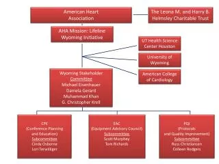
American Heart Association Guidelines for CPR 2015. Wanida Chongarunngamsang , MD. Faculty of Srinakarinwirot University. American Heart Association Guidelines for CPR 2015. BLS ACLS Pulseless Arrest Bradycardia Tachycardia Post cardiac arrest care. Chains of Survival.

E N D
American Heart Association Guidelines for CPR 2015 WanidaChongarunngamsang, MD. Faculty of Srinakarinwirot University
American Heart Association Guidelines for CPR 2015 • BLS • ACLS • Pulseless Arrest • Bradycardia • Tachycardia • Post cardiac arrest care
Chains of Survival 2015 (New): Separate Chains of Survival (Figure 4) have been recommended that identify the different pathways of care for patients who experience cardiac arrest in the hospital as distinct from out-of-hospital settings
“Chain of Survival”in-hospital cardiac arrest (IHCA) • Surveillance for cardiac arrest • Activate code (multidisciplinary team) • Initiate CPR by professional providers • Early defibrillation • Integrated post cardiac arrest care
“Chain of Survival”out-of-hospital cardiac arrest (OHCA) • Immediate recognition of cardiac arrest and activation of the emergency response system • Early CPR that emphasizes chest compressions • Rapid defibrillation if indicated • Effective advanced life support • Integrated post cardiac arrest care
Basic Life Support • Used for patients with life-threatening illness or injury before the patient can be given full medical care. • Generally used in the pre-hospital setting, and can be provided without medical equipment. • Generally does not include the use of drugs or invasive skills. .
30:2 x 5 cycle unresponsesive Call for help and AED Pulse :breathing 5-6 sec Breathing and pulse No pulse : CPR
The BLS Survey includes four steps: The BLS survey is the starting point for all ACLS management • Check for a response • Call for help and to bring an AED • Check circulation • Check rhythm
The Primary Assessment The Primary Assessment uses the ABCDE model • Airway – Use the least advanced airway possible to maintain the airway and oxygenation • Breathing – Monitor tube placement and oxygenation using waveform capnography • Circulation – Medications, CPR, fluids and defibrillation • Differential Diagnosis –treat reversible causes • Disability- neurological assessment “AVPU”(Alert, Voice, Painful, Unresponsive) • Exposure- looking for signs of trauma, bleeding, burns, or medical alert bracelets
The Secondary Assessment • The secondary assessment includes a search for underlying causes for the emergency and if possible a focused medical history “SAMPLE’ • (S)Signs and symptoms • (A)Allergies • (M)Medications • (P)Past Illnesses • (L)Last Oral Intake • (E)Events Leading Up To Present Illness
Check pulse :carotid artery • Start Chest compression if no definite pulse within 10 seconds
Chest compression Push hard, Put fast กดลึก-- ปล่อยสุด -- อย่าหยุด-- กดบ่อย
Chest compression กดลึก 5 cm (2 inches) Full chest recoid
Minimal interruption • หลัง defibrillation หรือ shock ให้กดหน้าอกต่อ ไม่ต้องคลำชีพจร • minimize the frequency and duration of interruptions in compressions • CPR without an advanced airway, goal of a chest compression fraction as high as possible, with a target of at least 60%.
Chest compression กดต่อเนื่องด้วยความเร็ว 100-120 ครั้งต่อนาที
Airway Head tilt Chin lift
AED (Automated External Defibrillator) • เปิดเครื่อง AED • ติด paddle ตามรูป • เครื่องจะทำการวิเคราะห์ว่าให้ shock ได้หรือไม่ • ถ้าเครื่องให้ shock ได้ให้กดปุ่มshock ที่เครื่อง
AED (Automated External Defibrillator) กดปุ่มเครื่องเปิด และหมุนปุ่มAED on AED ON
AED (Automated External Defibrillator) ติด pad ที่ sternum /apex
AED (Automated External Defibrillator) ต่อสาย electrode pad ต่อเข้ากับelectrode cable ของตัวเครื่อง
AED (Automated External Defibrillator) • เครื่องวิเคราะห์คลื่นไฟฟ้าหัวใจเมื่อเครื่องวิเคราะห์จะรายงานขึ้นบนจอภาพว่าเป็นคลื่นไฟฟ้าหัวใจแบบไหนและจะแนะนำว่าให้ทำ defibrillation ถ้าคลื่นไฟฟ้าหัวใจเป็นชนิดVF หรือVT • ห้ามสัมผัสผู้ป่วยเนื่องจากเครื่องจะอ่าน EKG ผิด • ถ้า EKG เป็นชนิดVF หรือVT เครื่องจะให้ charge พลังงาน • ถ้า EKG เป็นชนิด asystole เครื่องจะให้ CPR ต่อไป 2 นาทีแล้วจะ analyze EKG ใหม่
AED (Automated External Defibrillator) กดเพื่อทำการ shock
Advanced Cardiovascular Life Support: ACLS • Pulseless Arrest • Bradycardia with Pulse • Tachycardia with Pulse
shock • Toxins • Tamponade • Tension PTX • Thrombosis (coronary) • Thrombosis (pulmonary) • Hypovolemia • Hypoxia • Hydrogen ions (acidosis) • Hyper/hypokalemia • Hypothermia Amiodarone 300 mg----150 mg 5 Hs, 5Ts Pulse/BP EtCO2>40 mmHg A-line wave form
Narrow regular 50-100 j Narrow irregular 120-200 j (mono 200j) Unstable Tachycardia Wide regular 100 j Wide irregular DF
Quantitative Waveform Capnography • Confirmation and monitoring ETT placement • Evaluating the effectiveness of chest compressions ETCO2 value is at least 10-20 mmHg. • Identification of ROSC • Failure to achieve an ETCO2of greater than 10 mm Hg by waveform capnography after 20 minutes of CPR decide to end resuscitative efforts but should not be used in isolation
CPR Quality • Quantitative waveform capnography • If Petco2<10 mm Hg, attempt to improve CPR quality • Intra-arterial pressure • If relaxation phase (diastolic) pressure <20 mm Hg, attempt to improve CPR quality
SBP >90 mmHg MAP>65 mmHg BT 32C-36C at least 24 hr
Pulseless electrical activity(PEA) NO PULSE
New and Updated RecommendationsCPRGuideline 2015 • Separate Chain of Survival • Chest compressions at a rate of 100 to 120/min : extremely rapid compression rates with inadequate compression depth • Chest compressions at a depth of at least 2 inches or 5 cm for an average adult, while avoiding excessive chest compression depths (greater than 2.4 inches [6 cm])
New and Updated RecommendationsCPRGuideline 2015 • Delivery 1 breath every 6 seconds (10 /min) while continuous chest compression with advance airway • Vasopressin was removed from the ACLS Cardiac Arrest Algorithm • Nonshockable rhythm ,administer epinephrine as soon as feasible (IV/IO/ET) • Targeted temperature management 32C to 36C in 24 hr • The routine prehospital cooling of patients with rapid infusion of cold IV fluids after ROSC is not recommended