Download
1 / 49
510 likes | 750 Vues
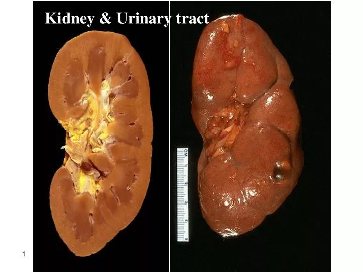
Kidney & Urinary tract. CLINICAL MANIFESTATIONS OF RENAL DISEASES. 1-Azotemia refers to an elevation of blood urea nitrogen(BUN) and creatinine levels It is largely related to a decreased glomerular filtration rate (GFR ). 2-uremia

E N D
CLINICAL MANIFESTATIONS OF RENAL DISEASES 1-Azotemia refers to an elevation of blood urea nitrogen(BUN) and creatinine levels It is largely related to a decreased glomerular filtration rate (GFR). 2-uremia when azotemia progresses to clinical manifestations and systemic biochemical abnormalities. Uremia is characterized by: 1- failure of renal excretory function. 2- metabolic and endocrine alterations
3- 2ry gastrointestinal manifestations(e.g., uremic gastroenteritis). 4- 2ry neuromuscular manifestations(e.g., peripheral neuropathy). 5- 2ry cardiovascular manifestations(e.g., uremic fibrinouspericarditis).
The major renal syndromes 1-Acute nephritic syndrome: it is a glomerular syndrome characterized by: 1- acute onset . 2- gross hematuria. 3- mild to moderate proteinuria (< 3.5 gm of protein/day in adults) 4- azotemia. 5- edema. 6- hypertension.
2-Nephrotic syndrome it is a glomerular syndrome characterized by: 1- heavy proteinuria (excretion of >3.5 gm of protein/day in adults) 2- hypoalbuminemia 3- severe edema 4- hyperlipidemia 5- lipiduria (lipid in the urine).
3-Asymptomatic hematuria or proteinuria is usually a manifestation of mild glomerular abnormalities. 4-Rapidly progressive glomerulonephritis It results in loss of renal function in a few days or weeks It is manifested by : 1-microscopic hematuria. 2-dysmorphic red blood cells and red blood cell casts in the urine sediment. 3-mild-moderate proteinuria
5-Acute renal failure is dominated by oliguria or anuria (no urine flow). recent onset of azotemia. It can result from : 1-glomerular injury (such as crescentic glomerulonephritis). 2-interstitial injury. 3-vascular injury (such as thrombotic microangiopathy). 4-acute tubular necrosis.
6- Chronic renal failure It is characterized by prolonged symptoms and signs of uremia. It is the end result of all chronic renal diseases . 7- Urinary tract infection It is characterized by bacteriuria and pyuria (bacteria and leukocytes in the urine). The infection may be symptomatic or asymptomatic. Types : 1- pyelonephritis (affection of the kidney ). 2- cystitis (affection of the bladder).
8-Nephrolithiasis Renal stones. It is manifested by: 1-renal colic. 2-hematuria. 3-recurrent stone formation.
GLOMERULAR DISEASES chronic glomerulonephritis is one of the most common causes of chronic kidney disease in humans. the glomerulus consists of an anastomosing network of capillaries invested by two layers of epithelium. The visceral epithelium (podocytes) is an intrinsic part of the capillary wall. the parietal epithelium lines Bowman space (urinary space), the cavity in which plasma ultrafiltrate first collects. The glomerular capillary wall is the filtration unit and consists of : 1-A thin layer of fenestrated endothelial cells, each fenestra 70 to 100 nm in diameter. 2-A glomerular basement membrane (GBM).
The capillary basement membrane • consists of : • 1- a thick electron-dense central layer (lamina densa) • 2- thinner and electron-lucent peripheral layers (lamina rarainterna and lamina raraexterna ). • The GBM consists of collagen (mostly type IV), laminin, polyanionicproteoglycans, fibronectin, and several other glycoproteins. • 3-The visceral epithelial cells (podocytes), structurally complex cells that possess interdigitating processes embedded in and adherent to the lamina raraexterna of the basement membrane.
Adjacent foot processes are separated by 20- to 30-nm-wide filtration slits which are bridged by a thin slit diaphragm composed in large part of nephrin. • 4-Supportive cells (mesangial cells) lying between the capillaries. • Basement membrane-like mesangial matrix forms a meshwork through which the mesangial cells are scattered.
Normal glomerulus by LM. The glomerular capillary loops are thin and delicate. Endothelial and mesangial cells are normal in number. The surrounding tubules are normal.
EM-GLOMERULUSCL-capillary lumen, End-endothelium, US-urinary space, B-basement membrane, Ep-epithelial cell, Mes-mesangial cell, Fp-foot process. Fp
The major characteristics of glomerular filtration 1- an extraordinarily high permeability to water and small solutes 2- an almost complete impermeability to molecules of the size and molecular charge of albumin (size: 3.6 nm radius; 70,000 kD). The selective permeability discriminates among protein molecules depending on: 1- their size (the larger the less permeable), 2- their charge (the more cationic the more permeable). 3-their configuration. Nephrin and its associated proteins, including podocin, have a crucial role in maintaining the selective permeability of the glomerular filtration barrier.
Pathogenesis of Glomerular Diseases Antibody-associated (1) injury resulting from deposition of soluble circulating Ag-Ab complexes in the glomerulus. (2) injury by Abs reacting in situ within the glomerulus. )3) Abs directed against glomerular cell components. 1-Nephritis Caused by Circulating Immune Complexes The antigen is not of glomerular origin. 1- endogenous as in the GN associated with SLE. 2- exogenous as in the GN that follows certain bacterial (streptococcal), viral (hepatitis B), parasitic (Plasmodium falciparum malaria), and spirochetal(Treponemapallidum) infections
antigen-antibody complexes are formed in situ or in the circulation and are then trapped in the glomeruli → activation of complement and the recruitment of leukocytes → injury. the glomerular lesions usually consist of leukocytic infiltration (exudation) into glomeruli and variable proliferation of endothelial, mesangial, and parietal epithelial cells. Electron microscopy reveals the immune complexes as electron-dense deposits or clumps that lie at one of three sites: 1-in the mesangium. 2-between the endothelial cells and the GBM (subendothelial deposits). 3-between the outer surface of the GBM and the podocytes (subepithelial deposits).
IF-Granular deposition of immune complexes characteristic of circulating and in situ immune complex deposition Deposits may be located at more than one site. The presence of Igs and complement in these deposits can be demonstrated by immunofluorescence microscopy. The pattern of immune complex deposition is helpful in distinguishing various types of GN
2-Nephritis Caused by In Situ Immune Complexes antibodies in this form of injury react directly with fixed or planted antigens in the glomerulus. Planted antigens include: 1- DNA. 2- bacterial products 3-large aggregated proteins (e.g., aggregated IgG), which deposit in the mesangium because of their size 4- immune complexes themselves because they continue to have reactive sites for further interactions with free antibody, free antigen, or complement.
3-Anti-Glomerular Basement Membrane (GBM) Antibody Glomerulonephritis Classic anti-GBM antibody GN (less than 1% of human GN cases). Abs are directed against fixed antigens in the GBM. Deposition of these antibodies creates a linearpattern of staining when the bound antibodies are visualized with IF microscopy.
The Nephrotic Syndrome The nephrotic syndrome refers to a clinical complex that includes the following: (1) massive proteinuriawith daily protein loss in the urine of 3.5 gm or more in adults. (2) hypoalbuminemiawith plasma albumin levels less than 3 gm/dL. (3) generalized edema (4) hyperlipidemia and lipiduria. (5) little or no azotemia, hematuria, or hypertension.
B-Systemic Diseases with Renal Manifestations: Diabetes mellitus: Amyloidosis Systemic lupus erythematosus drugs (gold, penicillamine, "street heroin") Infections (malaria, syphilis, hepatitis B, HIV) Malignancy (carcinoma, melanoma) Miscellaneous (e.g bee-sting allergy)
Minimal-Change Disease (Lipoid Nephrosis( This relatively benign disorder. The most frequent cause of the nephrotic syndrome in children (ages 1-7 years). It is characterized by glomeruli that have a normal appearance by LM but show diffuse effacement of podocytes by the EM. Pathogenesis: still not clear. Based on some experimental studies, the proteinuria has been attributed to a T-cell derived factor that causes podocyte damage and effacement of foot processes. neither the nature of such a putative factor nor a causal role of T cells is established in the human disease.
Morphology LM the glomeruli in minimal change disease appear normal. The cells of the proximal convoluted tubules are often heavily laden with protein droplets and lipids but this is secondary to tubular reabsorption of the lipoproteins passing through the diseased glomeruli (lipoid nephrosis). EM the GBM appears normal. The only obvious glomerular abnormality is the uniform and diffuse effacement of the foot processes of the podocytes . The cytoplasm of the podocytes thus appears flattened over the external aspect of the GBM obliterating the network of arcades between the podocytes and the GBM. There are also epithelial cell vacuolization microvillus formation and occasional focal detachments
MCD-EMthe capillary loop in the lower half contains two electron dense RBC's. Fenestrated endothelium is present and the BM is normal. The overlying epithelial cell foot processes are fused (arrows).
Clinical Course insidious development of the nephrotic syndrome in an otherwise healthy child. There is no hypertension. renal function is preserved in most individuals. selective proteinuria (the protein loss is usually confined to albumin ) The prognosisis good. When the changes in the podocytes reverse (e.g., in response to corticosteroids) the proteinuria remits More than 90% of cases respond to a short course of corticosteroid therapy. proteinuria recurs in more than 2/3 of the initial responders some of whom become steroid dependent < 5% develop chronic renal failure after 25 years and it is likely that most persons in this subgroup had nephrotic syndrome caused by FSGS not detected by biopsy. Adults with minimal change disease also respond to steroid therapy but the response is slower and relapses are more common.
Focal and Segmental Glomerulosclerosis characterized histologically by sclerosis affecting some but not all glomeruli (focal involvement) and involving only segments of each affected glomerulus. This histologic picture is often associated with the nephrotic syndrome. It can occur : (1)in association with other known conditions as AIDS or heroin abuse (HIV or heroin nephropathy). (2) as a secondary event in other forms of GN (e.gIgA nephropathy).
(3) as a maladaptation after nephron loss. (4) in inherited or congenital forms resulting from mutations affecting cytoskeletal or related proteins expressed in podocytes (e.g., nephrin). (5) as a primary disease( 20% to 30% of all cases of the nephrotic syndrome) . At least 50% of individuals with FSGS develop end-stage renal failure within 10 years of diagnosis. Adults do worse than children
Pathogenesis unknown . injury to the podocytes is thought to represent the initiating event of primary FSGS. permeability-increasing factors produced by lymphocytes. The deposition of hyaline masses in the glomeruli represents the entrapment of plasma proteins and lipids in foci of injury where sclerosis develops. IgM and complement proteins commonly seen in the lesion are also believed to result from nonspecific entrapment in damaged glomeruli. The recurrence of proteinuria after renal allografts transplantation sometimes within 24 hours of transplantation supports the idea that a circulating mediator is the cause of the damage to podocytes .
Morphology The disease is "focal" and initially affects only the juxtamedullaryglomeruli. With progression eventually all levels of the cortex are affected. LM-FSGS is characterized by lesions occurring in some tufts within a glomerulus and sparing of the others ( "segmental"). The affected glomeruli exhibit increased mesangial matrix, obliterated capillary lumens, and deposition of hyaline masses (hyalinosis) and lipid droplets. progression of the disease leads to global sclerosis of the glomeruli (global sclerosis) with pronounced tubular atrophy and interstitial fibrosis
focal and segmental glomerulosclerosis (PAS stain).a mass of scarred, obliterated capillary lumens with accumulations of matrix material
IF microscopy nonspecific trapping of Igs usually IgM, and complement in the areas of hyalinosis. EM the podocytes exhibit effacement of foot processes as in MCD. Clinical Course Poor responses to corticosteroid therapy. about 50% of individuals suffer renal failure after 10 years.
Collapsing glomerulopathy It is a morphologic variant of FSGS. It carries a particularly poor prognosis. It is characterized by collapse of the entire glomerular tuft and podocyte hyperplasia. It may be : 1-idiopathic . 2-associated with HIV infection. 3-drug-induced toxicities.
Membranous Glomerulonephritis ( MGN) It is slowly progressive disease. most common between 30 -50 years of age. It is characterized morphologically by the presence of subepithelialIg-containing deposits along the GBM. Membranous glomerulonephritis : 1-Idiopathic (85% of cases). 2-Secondary
secondary to other disorders including: (1) infections (HBV, syphilis,schistosomiasis, malaria). (2) malignant tumors (carcinoma of the lung and colon and melanoma). (3) autoimmune diseases as SLE . (4) exposure to inorganic salts (gold, mercury). (5) drugs (penicillamine, captopril,NSAID).
Pathogenesis Membranous GN is a form of chronic immune complex nephritis. circulating complexes of 1- exogenous (e.g., hepatitis B virus) . 2- endogenous (DNA in SLE) antigen . Morphology LM the basic change appears to be diffuse thickening of the GBM .
LM- membranous glomerulonephritis in which the capillary loops are thickened and prominent, but the cellularity is not increased
Membranous nephropathy. A, Diffuse thickening of the glomerular basement membrane. B, Schematic diagram illustrating subepithelial deposits, effacement of foot processes, and the presence of "spikes" of basement membrane material between the immune deposits.
A silver stain of the glomerulus highlights the proteinaceous basement membranes in black. There are characteristic "spikes" seen with membranous glomerulonephritis seen here in which the black basement membrane material appears as projections around the capillary loops.
IF • diffuse granular deposits of immunoglobulins and complement along the GBM . • mainly IgG and complement. • EM • subepithelial deposits → thickening of the GBM and are separated from each other by small, spikelike protrusions of GBM matrix that form in reaction to the deposits ("spike and dome" pattern). • As the disease progresses, these spikes close over the deposits, incorporating them into the GBM. • the podocytes showeffacement of foot processes.
MGNIF-deposits of mainly IgG and complement collect in the basement membrane and appear in a diffuse granular pattern
EM-the darker electron dense immune deposits are seen scattered within the thickened basement membrane.
Clinical Course insidious development of the nephrotic syndrome, usually without antecedent illness. some individuals with membranous nephropathy may have lesser degrees of proteinuria rather than the full-blown nephrotic syndrome. the proteinuria is nonselective. no response to corticosteroid therapy. Membranous nephropathy follows a variable and often indolent course. Overall proteinuria persists in over 60% of cases. ~ 40% suffer progressive disease terminating in renal failure after 2 to 20 years. 10%-30% have a more benign course with partial or complete remission of proteinuria.