Download
1 / 114
1.14k likes | 1.33k Vues
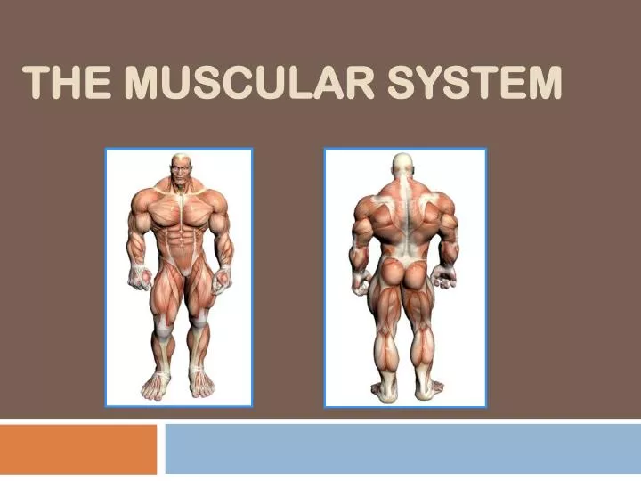
The Muscular System. Objectives. Indicate primary functions of muscles Identify and distinguish between three types of muscle in terms of location, structure, components, appearance, purpose Describe shapes associated with muscle, list an example of each. The Muscular System.

E N D
Objectives • Indicate primary functions of muscles • Identify and distinguish between three types of muscle in terms of location, structure, components, appearance, purpose • Describe shapes associated with muscle, list an example of each
The Muscular System • Muscles are responsible for all movement of the body, support and strengthen the skeletal frame by connecting to bone, provide heat as a by-product • There are three basic types of muscle • Skeletal • Cardiac • Smooth
Info About Muscles • Only body tissue able to contract • Movement created by flexingand extendingjoints • Body energyconverters (many muscle cells contain many mitochondria)
Skeletal Cardiac Smooth Three types of muscle
Characteristics of Muscle • Skeletal and smooth muscle are elongated • Muscle cell diff. than other “typical” cells • Size (much bigger) • Skeletal multinucleated
Characteristics of Muscle • Contraction of a muscle is due to movement of microfilaments (protein fibers) • All muscles share some terminology • Prefixes myo and mys refer to muscle • Prefix sarco refers to flesh
Skeletal Muscle • Most are attached by tendons to bones • Cells have more than one nucleus (multinucleated) • Striated- have stripes, banding • Voluntary- subject to conscious control • Tendons are mostly made of collagen fibers • Found in the limbs
Structure of skeletal muscle • Each cell (fibre) is long and cylindrical • Muscle fibres are multi-nucleated • Typically 50-60mm in diameter, and up to 10cm long • The contractile elements ofskeletal muscle cells aremyofibrils
Skeletal muscle - Summary • Voluntary movement of skeletal parts • Spans joints and attached to skeleton • Multi-nucleated, striated, cylindrical fibres
Smooth Muscle • No striations • Spindle shaped • Single nucleus • Involuntary- no conscious control • Found mainly in the walls of hollow organs
Smooth muscle • Lines walls of viscera • Found in longitudinal or circular arrangement • Alternate contraction of circular & longitudinal muscle in the intestine leads to peristalsis
Structure of smooth muscle • Spindle shaped uni-nucleated cells • Striations not observed • Actin and myosin filaments are present( protein fibers)
Smooth muscle - Summary • Found in walls of hollow internal organs • Involuntary movement of internal organs • Elongated, spindle shaped fibre with single nucleus
Cardiac Muscle • Striations • Branching cells • Involuntary • Found only in the heart • Usually has a single nucleus, but can have more than one
Cardiac muscle • Main muscle of heart • Pumping mass of heart • Critical in humans • Heart muscle cells behave as one unit • Heart always contracts to it’s full extent
Structure of cardiac muscle • Cardiac muscle cells (fibres) are short, branched and interconnected • Cells are striated & usually have 1 nucleus • Adjacent cardiac cells are joined via electrical synapses (gap junctions) • These gap junctions appear as dark lines and are called intercalated discs
Cardiac muscle - Summary • Found in the heart • Involuntary rhythmic contraction • Branched, striated fibre with single nucleus and intercalated discs
Shapes of Muscles • Triangular- shoulder, neck • Spindle- arms, legs • Flat- diaphragm, forehead • Circular- mouth, anus
Objectives • Distinguish between origin and insertion • Explain the difference between twitch and tetanus • Explain the difference between isotonic and isometric contractions. Provide an example of each • List the muscles associated with the muscles of the head and neck
Objectives cont. • List, identify and describe movement associated with the 6 muscles of the face • List, identify and describe movement associated with 2 muscles involved in mastication • List, identify and describe movement associated with primary muscle of the neck
How are Muscles Attached to Bone? • Origin-attachment to a movable bone • Insertion- attachment to an immovable bone • Muscles are always attached to at least 2 points • Movement is attained due to a muscle moving an attached bone
Insertion Origin Muscle Attachments
Types of Responses • Twitch- • A single brief contraction • Not a normal muscle function • Tetanus • One contraction immediately followed by another • Muscle never completely returns to a relaxed state • Effects are compounded • “Disease” caused by bacteria entering dirty wound complication includes severe cramping due to cont. tetanic contractions
Where Does the Energy Come From? • Energy is stored in the muscles in the form of ATP • ATP comes from the breakdown of glucose during Cellular Respiration • This all happens in the Mitochondria of the cell • When a muscle is fatigued (tired) it is unable to contract because of lack of Oxygen
Exercise and Muscles • Isotonic- muscles shorten and movement occurs ( most normal exercise) • Isometric- tension in muscles increases, no movement occurs (pushing one hand against the other, pushing against a wall); energy is still used! • Athletes use this to strengthen joints, burn calories while minimizing enlargement of muscle mass
The Skeletal MusclesThere are about 650 muscles in the human body. They enable us to move, maintain posture and generate heat. In this section we will only study a sample of the major muscles.
Naming of Muscles • Each named in terms of appearance, location, action or relation to other structures • Latin or Greek in origin
Muscles of the Head and Neck • Includes muscles that provide facial expressions, muscles involved in chewing and muscles in the neck that aid in movement • Muscles of Facial Expression • Muscles of Mastication • Neck Muscle
Muscles of Facial Expression • Several muscles act on facial structures to provide manipulation of facial movement • Frontalis • Occipitalis • Orbicularis • Orbicularis oris • Buccinatior • Zygomaticus
Muscles of Facial Expression • Frontalis • Forehead • Raises the eyebrows • Occipitalis • Located on lower back of head • Pulls scalp backward
Muscles of Facial Expression • Orbicularis oculi • Surrounds each eye • Closes eye, squinches eye • Orbicularis oris • Surrounds mouth • Closes mouth
Muscles of Facial Expression • Buccinator • In conjuction w/orbicularis oculi and orbicularis oris enables mouth to pucker • Also flattens cheeks to enable whistling/blowing
Muscles of Facial Expression • Zygomaticus • Corner of mouth • Smiling
Muscles of Mastication • Two major muscles involved in chewing (mastication) • Masseter • Extends from zygomatic process of temporal bone to the mandible • Closes mouth; can be felt on inside jaw during chewing • Temporalis • Occupies most of temporal bone • Closes mouth
Neck Muscle • Sternocleidomastoid • Anterior and posterior sides of neck • From anterior view, origin of the two sternocleidomastoids forms a “V” at base of neck • Allows movement of the head; contraction of both muscles will flex the head, contraction of one will cause rotation
Neck Muscle - Sternocleidomastoid Allows head movement
Objectives • Define trapezius in terms of description, location, movement • List 4 muscles associated with pectoral girdle and their overall purpose • Define pectoralis major, latissimus dorsi and deltoid in terms of description, location, movement and any abbreviations each may have
Objectives • List purpose of rotator cuff muscles • Define triceps and biceps in terms of description, location, number of origins, movement • Differentiate between posterior and anterior movement of wrist and fingers
Upper Limb Muscles • Include those that attach and support the pectoral girdle, upper limb and those those move the arm, forearm and hand • Muscles of Pectoral Girdle • Muscles that Move the Arm • Muscles that Move the Forearm • Muscles that move the Fingers
Upper-Limb Muscles • Muscles of the Pectoral Girdle • Connection of girdle to thorax supported by group of muscles • Trapezius • Diamond shaped, extends from base of the skull to the middle of the back • Contraction causes scapula to move up and down; rotates shoulder
Upper Limb Muscles • Levator scapulae • Origin:1st four cervical vertebrae • Rhomboids • Origin: 7th cervical and 1st five thoracic vertebrae • Serratus anterior • Origin: 1st eight ribs • Pectoralis minor • Origin: Ribs 3-5 • Hold together scapula when arm muscles contract
Upper Limb Muscles Levator Scapulae Rhomboid