Download
1 / 27
270 likes | 301 Vues
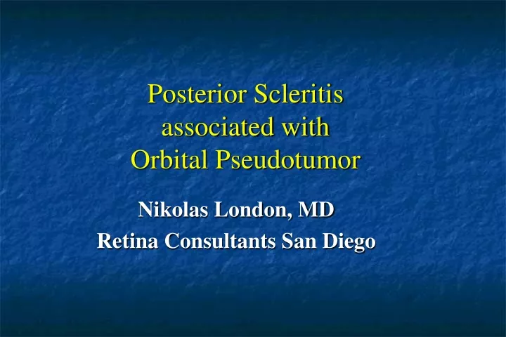
Posterior Scleritis associated with Orbital Pseudotumor. Nikolas London, MD Retina Consultants San Diego. Ocular History. 34-year-old man with 2 months of headache , progressive proptosis , pain , redness , and decreased vision in his right eye

E N D
Posterior Scleritis associated withOrbital Pseudotumor Nikolas London, MD Retina Consultants San Diego
OcularHistory • 34-year-old man with 2 monthsofheadache, progressive proptosis, pain, redness, anddecreasedvision in hisrighteye • HPI: Pred Forte andscopolaminefor NGAU x 4 weeks • POHx: none • PMH: • Mitralvalveprolapse • Mental illness: self-described“not right in head” • Jawsurgery 1994 • ALL: mushrooms, mayonnaise, anabolicsteroids • SH: NC • ROS: pan-negative
First Presentation • VA: bare CF OD, 20/25 OS • Pupil: + RAPD OS byreverse • IOP: 15 OU • Hertel: 5mm proptosis OD • SLE OD: 2+ conjunctival injection, 1+ AC andanteriorvitreous cell
First Presentation • Funduscopy • large amelanonic mass superior to the optic nerve head causing • retinal folds and • obscuration of the optic nerve head.
First Presentation • Fluoresceineangiography • Early widefield angiogram of the right eye • retinal distortion and folds. • later frames: progressive stippled hyperfluorescence of the mass • prominent leakage from the optic nerve head
First Presentation • Fluoresceineangiography • Late frame widefieldangiogram of the right eye • leakage from the mass and optic nerve head • inferior peripheral nonperfusion and adjacent vascular leakage.
First Presentation • US • vertical axial B-scan ultrasound • thickening of the posterior wall complex with sub-Tenon’s fluid (T-sign) • shallow inferior retinal detachment
First Presentation • periorbitaledema • erythema • mild exotropia and proptosis.
First Presentation • Imaging of the right orbit • 2.7 x 1.8 x 3.3 cm soft tissue mass • involving the sclera with deformation of the posterior globe. • Pseudotumor orbitae (?)
Diagnosis • Posterior scleritis • Associated to pseudotumor orbitae • workupforinfectiousandinflammatoryetiologies • senttoOculoplasticsforevaluationtoconsiderbiopsyandrule out lymphoma. • biopsy was refusedbecausequiterisky
Laboratory Data Quantiferon gold negative FTA-ABS NR RPR NR ACE 21 C-ANCA negative P-ANCA negative X-ANCA negative ANA negative CXR wnl ESR 25 CRP 1.10 Chem-7 wnl CBCD mild anemia Hgb/Hct 12/36
Treatment and Follow-Up • after infectiousetiologieswereruled out he was started on prednisone (60mg/day, 2 weeks) • then reduction by 20 mg/week for 3 weeks, staying on 10 mg/day for several weeks • rapid improvementofhissymptomsandexamination in between 1 week • dramatic reduction in periorbital edema, erythema, propsosis, head tilt, and exotropia
Follow-Up1 week after beginoftreatment • dramatic reduction in size of the subretinal mass • residual RPE changes • mild horizontal retinal striae in the superior macula. • SD OCT: mild inner retinal distortion and subretinal fluid. • Imaging: substantially smaller scleral mass with less distortion of the posterior wall of the globe.
Final Diagnosis • Posterior scleritis • associated with idiopathic orbital pseudotumor • rapid resolution with oral corticosteroids
Conclusion • Posterior scleritis is a rare manifestationof orbital pseudotumor • Other diagnoses, includingtuberculosis, lymphoma, systemiclupus erythematosus, syphilis, and sarcoidosis shouldbeconsidered