Download
1 / 6
330 likes | 1.72k Vues
Cranial cavity. Internal surface of vault of skull,show sagital groove in median plane. Floor of cranial cavity is formed by upper surface of base of skull, it’s divided into 3 fossa: 1- ant. Carnial fossa 2- middle cranial fossa 3- post. Cranial fossa. Anterior Cranial fossa:.

E N D
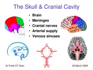
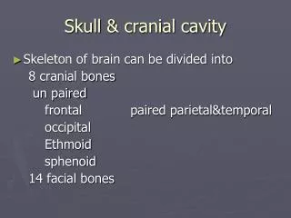
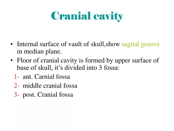
Cranial cavity • Internal surface of vault of skull,show sagital groove in median plane. • Floor of cranial cavity is formed by upper surface of base of skull, it’s divided into 3 fossa: 1- ant. Carnial fossa 2- middle cranial fossa 3- post. Cranial fossa
Anterior Cranial fossa: It’s floor formed by 3 bones : 1) Ethmoid bone 2) orbital plate of frontal bone which form roof of orbit 3) sphenoid bone • It’s present crista galli which median projection of ethmoid bone • On each side of crista galli there is cribiform plate of ethmoid • foramen cecum • Frontal crest
middle cranial fossa • It consists of middle narrow part & 2 expanded lateral part • Median part is formed by body of sphenoid which called sella turcica & includes the following: • Dorsum sallae • Hypophyseal fossa. • Tuberculum sallae • Medial end of lesser wing sphenoid projects ant. Glenoid process. • Post. Glenoid process at lateral end • sup. Orbital fissure lies between greater&lesser wing of sphenoid.
post. Cranial fossa • Jugualr foramen: Foramen magnum which contain lower end of medulla oblingnata, • it’s the biggest foramen • Which transmit vertebral A. • Hypoglossal canal • It’s found between jugular tubercle which which elevation between jugular foramen & foramen magnum • Transmit hypoglossal N. • Below foramen magnum there is internal occipital crest to reach internal occipital protuberance.
ANY QUESTION ????? THANK YOU