Download
1 / 18
190 likes | 506 Vues
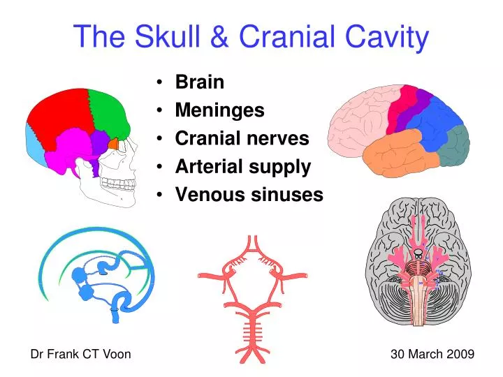
The Skull & Cranial Cavity. Brain Meninges Cranial nerves Arterial supply Venous sinuses. Dr Frank CT Voon. 30 March 2009. The Neurocranium. Frontal. Parietal. Occipital. Ethmoid. Temporal. Sphenoid. Tip: PETS OF or FPOETS. The Cranium.

E N D
The Skull & Cranial Cavity • Brain • Meninges • Cranial nerves • Arterial supply • Venous sinuses Dr Frank CT Voon 30 March 2009
The Neurocranium Frontal Parietal Occipital Ethmoid Temporal Sphenoid • Tip: PETS OF or FPOETS.
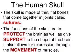
The Cranium • The cranium (skull) is the skeleton of the head. • It consists of a neurocranium and a viscerocranium. • The neurocranium is also known as the cranial vault. • The viscerocranium is also known as the facial skeleton.
The Neurocranium • The neurocranium (cranial vault) has a roof and a floor. • The roof (calvaria or skull cap) is shaped like a dome. • The basicranium (cranial base) forms the floor. • It encloses the cranial cavity.
The Neurocranium • It is formed by 8 bones, the frontal, parietal, occipital, temporal, ethmoidal and sphenoidal bones. • The frontal, occipital, ethmoidaland sphenoidalbones are single and thus are in the midline. • The parietal and temporal bones are bilateral and hence are paired. • The fibrous joints between the bones are known as sutures.
Intramembranous ossification • The frontal, parietal and temporal bones are formed by intramembranous ossification and are known as flat bones. • Do note that these flat bones forming the calvaria are actually curved, with a convex external surface and a concave internal surface.
Endochondral ossification • The sphenoid, ethmoid and temporal bones are mainly formed by endochondral ossification and are known as irregular bones. • These bones form the cranial base. • The ethmoid is a part of both the neurocranium and viscerocranium.
The Brain and spinal cord • Cerebral hemispheres • Diencephalon • Midbrain • Pons and cerebellum • Medulla oblongata • Spinal cord
Sensory cortex Motor cortex Parietal lobe Frontal lobe Occipital lobe Temporal lobe
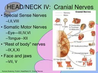
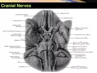
Olfactory Optic Oculomotor Trochlear Trigeminal Abducens Facial Vestibulocochlear Glossopharyngeal Vagus Accessory Hypoglossal The Cranial nerves
Foramina • Structures that pass through the foramina • Cranial nerves • Arteries - Internal carotid and vertebral • Veins – sigmoid sinus and beginning of internal jugular vein • Spinal cord – Foramen magnum
Circle of Willis Anterior communicating artery Anterior cerebral artery Perforating arteries Internal carotid artery Ophthalmic artery Middle cerebral artery Anterior choroidal artery Posterior communicating artery Posterior cerebral artery Superior cerebellar artery Basilar artery Pontine arteries Anterior inferior cerebellar artery Vertebral artery
Superior sagittal sinus Arachnoid granulations Emissary veins Skin Close tissue Aponeurosis Loose tissue Periosteum Outer table Diploe Inner table Dura mater Endosteum Arachnoid mater Subdural space Pia mater Gray matter Subarachnoid space White matter Falx cerebri Cerebrospinal fluid
Venous sinuses Superior Sagittal Sinus Inferior Sagittal Sinus Confluence Straight Sinus Internal occipital protruberance Superior Petrosal Sinus Occipital Sinus Sigmoid Cavernous Sinus Sinus Transverse Sinus Inferior Petrosal Sinus Jugular foramen Internal Jugular Vein
Skin Close subcutaneous tissue Aponeurosis Loose areolar tissue Periosteum Outer table Diploe Inner table Endosteum The Scalp and Cranial Cavity • Endosteal layer of dura mater • Menigeal layer of dura mater • Arachnoid mater • Pia mater • Brain • Grey matter • White matter • Ventricles
Meninges and spaces • Extradural space • Dura mater • Subdural space • Arachnoid mater • Subarachnoid space • Pia mater • Gray matter • White matter • Ventricles