Download
1 / 80
800 likes | 981 Vues
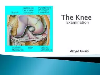
Chapter 10: The Knee. The bones that comprise the knee joint: Tibia Fibula Femur Patella There are two joints in the knee: Tibiofemoral joint Patellafemoral joint. Anatomy. Anatomy. The medial and lateral meniscus rest between the femur and tibia.

E N D
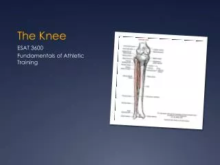
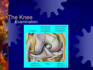
The bones that comprise the knee joint: Tibia Fibula Femur Patella There are two joints in the knee: Tibiofemoral joint Patellafemoral joint Anatomy
Anatomy • The medial and lateral meniscus rest between the femur and tibia. • They are responsible for shock absorption, improved bony correlation, joint lubrication, improved weight distribution, and decreased friction. • The patella guides the quadriceps, decreases friction during movement, and protects the femoral condyles.
Anatomy of the Knee • Knee is a hinge joint • Articulation (point of contact) • Consists of 3 bones • Stabilized by • Four major ligaments • Cartilage • Strong musculature • Knee is able to rotate
Cartilage • Ends of the tibia and femur • Covered/cushioned by • Pieces of tough cartilage tissue • Called menisci • Help to stabilize the knee joint • Without bones would rub & wear down quickly • Top of tibia • Flat like a tabletop • End of femur • Rounded (called condyles) • Without stabilization, femur would move a lot on the tibia
Cartilage-Menisci • Lateral & Medial • Thicker on sides • Thinner in the middle • Form a dish-shaped hollow • Attached to the top of the tibia • Provide a seat for the femoral condyles to sit in • Femur moves but will not roll off
Ligaments • 4 primary knee ligaments • Medial collateral (MCL) • Helps provide stability to inside of knee • Lateral collateral (LCL) • Helps provide stability to outside of knee • Anterior cruciate ligament (ACL) • Keeps tibia from moving forward on the femur • Posterior cruciate ligament (PCL) • Prevents tibia from moving backward on the femur • ACL & PCL • pass through the middle of the knee joint • Cross each other (i.ecruciate means “cross-shaped”)
Ligaments of the Knee • Anteriorcruciate ligament (ACL) • Posteriorcruciate ligament (PCL) • Medial collateral ligament (MCL) • Lateral collateral ligament (LCL)
Anatomy • Muscles of the Knee • Quadriceps muscles • Responsible for knee extension • Hamstring muscles • Responsible for knee flexion • Calf muscles • Assist in knee flexion • Other muscles that act at the knee • Sartorius • Popliteus • Plantaris • Gracilis
Muscles of the Knee • Provide • Movement • Stability • Primary muscles spanning the knee • Quadriceps group (perform knee extension) • Vastusmedialis, vastuslateralis, vastusintermedius & rectus femoris • Hamstring group (perform knee flexion) • Biceps femoris, semimembranosus & semitendinosus • Help prevent forward movement of the tibia on the femur • By the location of their attachments
Primary Muscles Spanning the Knee • Quadriceps group • Vastusmedialis, vastuslateralis, vastusintermedius & rectus femoris • Perform knee extension
Primary Muscles Spanning the Knee • Hamstring group • Biceps femoris, semimembranosus & semitendinosus • Perform knee flexion • Help prevent forward movement of the tibia on the femur • By the location of their attachments
Knee Alignment Concerns • Genuvalgum(knock-knees) • Genuvarum(bow legs) • Genurecurvatum(hyperextension) • Q-angle • Greater than 20 increases risk for injury • Leg length discrepancy
Preventing Knee Injuries • Ligament sprains • Most common injuries seen at the knee • Muscles provide stability to the knee • Help resist abnormal bony movement • Athletes should develop strength in the muscles (quads, hams, gastrocnemius/calf, hip abductors & hip adductors) • Gastrocnemius-heel raises • Some trainers & athletes use preventative knee braces • Designed to protect medial collateral ligament • Tearing can result from a blow to the lateral side
Treating Knee Injuries & Conditions • Knee is exposed to many forces • Makes it vulnerable to injuries • Especially the ligaments • Tendon & bone injuries also occur • Patella & menisci are subject to unique types of athletics-related injuries
Ligament Injuries • Ligament sprains of the knee • Can be • Mild • Moderate • Severe
Patellar Fracture • Signs & Symptoms: • Pain directly over bone • Slight to moderate swelling • Pain, especially in the first 30º of movement • Treatment: • Immobilize and refer to a physician for x-rays • Requires lengthy immobilization during recovery
Patellar Dislocation • Signs & Symptoms: • Moderate to extreme pain • Moderate swelling • Complete loss of ROM in knee • Obvious deformity laterally • Treatment: • Refer to physician for reduction • RICE therapy • Progressive strengthening program
Patella Dislocation • Patella forced to lateral aspect of the knee • Often occurs when the knee is bent and forced to twist inward • Signs & symptoms • Obvious deformity • Athlete is often in distress • EMS must be called • Unless team physician is present • only a physician should reduce a dislocated patella • Complications may result • Posterior aspect of patella may be injured further
Patella-Femoral Stress Syndrome • Signs & Symptoms: • Pain and tenderness in lateral aspect of patella • Slight swelling • Crepitus or popping with extension • Treatment: • RICE therapy • Closed kinetic chain exercises 0-40
Patellar Femoral Syndrome • Fancy name for a set of symptoms that include pain/discomfort around patella • Often caused by patellar tracking problems • Knee bends • Patella is grated across the femur • Causing cartilage on back of patella to soften or wear away • Known as chondromalacia
Chondromalacia • Signs & Symptoms: • Pain underneath the patella • Grinding or popping during motion • Slight chronic swelling • Special Tests: Clarke’s Sign • Treatment: • RICE Therapy • Quadriceps strengthening
Patellar Femoral SyndromeChondromalacia • Characterized by achiness around the patella • Especially with prolonged sitting in the same position • Athlete reports a grinding sensation with flexion/extension • Grinding can be felt by placing hand over knee
Osgood-Schlatter’s Disease • Signs & Symptoms: • Pain at insertion of patella tendon • Tenderness to palpation • Enlarged tibialtuberosity • Pain with jumping or running • Treatment: • RICE therapy • Decrease activity or cross-train
Osgood-Schlatter Disorder • Irritation at the site of the patellar tendon attachment • To front of the tibia • Called tibialturberosity • Repeated stress causes the patellar tendon to partially pull away from the bone • Called Osgood-Schlatter’s disorder
Osgood-Schlatter Disorder • Signs & symptoms • Discomfort of the knee • Swelling • Tenderness • Pain during activity • Possible bump below knee cap (bony growth at the top of the tibia) • Can remain even after symptoms have disappeared • Care • Restrict activity until resolved • Stationary bicycling • Use pain as a guide • Modify activities based on pain level reported by athlete • Ice before & after activity • Special pad made to fit over front of tibia • Often improves by age 16 or 17 (but known to last into early 20’s)
Patellar Tendinitis • Signs & Symptoms: • Pain in patella tendon or at inferior pole of patella • Pain increases with activity • Squeaking noise with motion • Slight swelling • Treatment: • Modality treatment • Ice or ice massage • Ultrasound
Muscle & Tendon Injuries • Patellar tendinitis • Overuse disorder • Characterized by quadriceps weakness • Tenderness over the patellar tendon • Minimal swelling • Condition is also called jumper's knee • Athletes that do lots of jumping often get this condition (basketball, volleyball) • Early stages • Athlete typically has pain after activity • Treatment • Trainer attempts to control inflammation • Apply ice • Modify athlete’s activity level • Restricting running & jumping • Rehabilitation program • Address any flexibility problems or weakness of the leg
Patella Tendon Rupture • Signs & Symptoms: • Extreme pain with an immediate drop in pain • Significant swelling • Window shade effect • Complete loss of knee extension • Previous history of chronic tendinitis • Treatment: • Surgical repair is the only treatment option • 6-8 months minimum recovery
Knee Dislocation • Signs & Symptoms: • Immediate pain that may decrease dramatically • Obvious deformity (usually anteriorly) • Significant swelling • Decreased blood flow and neural sensation • Treatment: • Splinted and transported to hospital immediately • Surgical intervention is often required for neurovascular and ligament repair
Knee Contusion • Signs & Symptoms: • Pain at affected site • Moderate swelling and discoloration • Loss of ROM • Decreased weight bearing • Treatment: • RICE therapy
Patella Injuries (Bursitis) Prepatellar bursitis is the inflammation of a small sac of fluid located in front of the kneecap. This inflammation can cause many problems in the knee.
Causes Bursitis is the inflammation of a bursa. The prepatellar bursa can become irritated and inflamed in a number of ways. A direct blow or a fall onto the knee can damage the bursa.
Meniscus Contusion • Signs & Symptoms: • Pain, especially at full extension • Loss of ROM in extension • Slight swelling • Treatment: • RICE therapy
Meniscus Tears • Signs & Symptoms: • Pain, especially when moved similarly to the mechanism of injury • Pain with full extension or flexion • Diffuse swelling in the joint (effusion) • Pain along the line of the joint • Sensation of locking or giving out • Clicking or popping sound with movement
Meniscal Injuries Meniscal injuries damage the cushioning tissue between the tibia and the femur, inside the knee joint, on both sides (medial and lateral) of the knee.
Causes They are highly vulnerable to injury from abrupt rotations of the knee while it is bearing weight, for example, when you turn to hit a tennis ball, rotating your thigh (femur) while leaving your foot stationary.
Meniscus Tears • Special Tests: • McMurray’s Test • Apley’s Compression Test • Bounce Home Test • Treatment: • Referral to a physician • Surgery is often required for full recovery. • RTP depends on surgical option selected.
ACL Sprain • Signs & Symptoms: • Pain in the joint • Athlete hears ‘pop’ at time of injury • Sense of looseness in joint, giving away, or shifting • Swelling that increases rapidly post-injury
Anterior Cruciate Ligament Injuries • Keeps tibia from moving forward on the femur • If ligament is injured • Athlete is often disabled • Complaining of the knee “giving away”, collapsing & popping • Most serious of all knee ligament injuries • Most frequently surgically reconstructed