Download

1 / 18
180 likes | 645 Vues
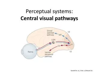
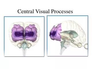
Corteza Visual Primaria (V1). Curva de afinación a la orientación. Estímulos óptimos y no óptimos. DA, Figs 1.5 & 2.11 . Parametrización del CR de las células simples con funciones de Gabor. DA, Fig 2.12 . Ajuste de los CR’s con Gabors. D Ringach, J Neurophysiol 88: 455-463, 2002

E N D
Curva de afinación a la orientación Estímulos óptimos y no óptimos DA, Figs 1.5 & 2.11
Parametrización del CR de las células simples con funciones de Gabor DA, Fig 2.12
Ajuste de los CR’s con Gabors D Ringach, J Neurophysiol 88: 455-463, 2002 http://jn.physiology.org/cgi/content/full/88/1/455
Distribución de CR’s n_x = |f| sigma_x n_y = |f| sigma_y D Ringach, Figs 4 & 5
Distribución de fases D Ringach, Fig 6
Predicciones de CR’s a partir de imágenes naturales ICA SC D Ringach, Figs 8, 9 & 10
Dinámica de los CR’s Tiempo óptimo (mseg) D Ringach, Fig 1
Otras propiedades de selectividad: disparidad f(s) = r_max/[1 + exp( s_{1/2}-s / (\Delta \sigma) )] DA, Fig 1.7
Células complejas (tipo 1) Nicholls, Fig 20.13
Modelo para el CR de células complejas Nicholls, Fig 20.15
Propiedades de los CR de células visuales desde la retina hasta V1 Nicholls, tabla 20.1
V1: capas, entradas y salidas Nicholls, Fig 20.7
Columnas de orientación Nicholls, Fig 21.3
Columnas de dominación ocular en primates Figure 23-16 KSJ, Fig 23.16
Columnas de dominación ocular Nicholls, Fig 21.1
Figure 23-17 Hiper-columnas KSJ, Fig 23.17