Download
1 / 31
670 likes | 1.42k Vues
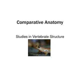
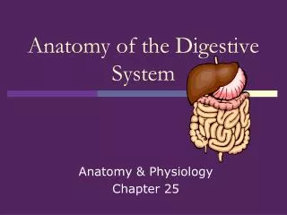
Comparative Anatomy Digestive System. Note Set 11 Chapter 13. Digestive System. Six major subdivisions Oral cavity Pharynx Esophagus Stomach Small & large intestine Rectum. Digestive System. Agnatha - straight digestive tube Coiled tube evolved with lengthening of tract.

E N D
Comparative AnatomyDigestive System Note Set 11 Chapter 13
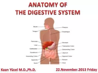
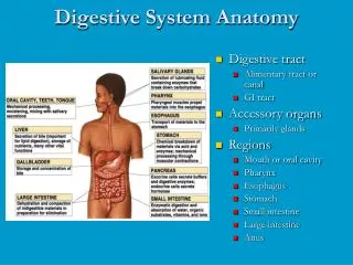
Digestive System Six major subdivisions • Oral cavity • Pharynx • Esophagus • Stomach • Small & large intestine • Rectum
Digestive System • Agnatha - straight digestive tube • Coiled tube evolved with lengthening of tract Figure 11.1: Simple to complex digestive systems.
Oral Cavity • Begins at mouth, ends at pharynx • Tongue in floor of cavity • Palate in roof of cavity • Primary palate • Secondary palate • Teeth Figure 11.2: Human oral cavity.
Palates • Primary palate in anamniotes- nasal passageways empty into oral cavity • Ex: Salamander • Secondary palate of amniotes- extends to pharyngeal cavity • Internal nares Figure 11.3: Oral cavity of amphibian (a) and mammal (b).
Teeth • On jaws normally • Cheeks in mammals form pocket • Acrodont teeth- fish and snakes • Bicuspid- amphibians • Tricuspid- lizards • Pleurodont teeth- snakes • Thecodont teeth- crocodilians Figure 11.5- Types of cusps. Figure 11.4- Cross section of jaw.
Jaw Teeth and Cheek • Used for storage- rodents and squirrels • Modified placoid scales- sharks • Polyhyodont- permanent replacement of teeth • Diphyodont- two sets of teeth • Monophyodont- one set of teeth
Bird Teeth • Egg caruncle- all egg layers • Not actual tooth • Structure epidermal, horny, keratinized • On tip of snout • To penetrate egg shell Figure 11.6: Egg caruncle of 15 day old owlet.
Reptilian Teeth • Egg tooth- lizards and snakes • Actual tooth • Upper jaw • To penetrate egg shell Figure 11.7: Monitor egg tooth..
Modifications of Snake Teeth • Aglyphous- no modifications for venom delivery • Solenoglyphous- retractable teeth, fangs • Proteroglyphous- fangs in front of mouth • Opisthoglyphous- fangs in back of mouth Figure 11.8: Position, cross and longitudinal sections of aglyphous (1), opisthoglyphous (2), and solenoglyphous (3) fangs.
Mammalian Teeth • Incisors • For cutting • Ex: elephant tusks • Canines • For piercing • Ex: walrus tusks • Premolars & Molars • To matriculate food • Diastema- space without teeth; e.g., no canines Figure 11.9: Mammalian teeth specializations.
Mammalian Teeth • Heterodont dentition • Other varieties • Homodont- all teeth the same • Bunodont- all teeth on single plain • Sectorial teeth – carnassials; e.g., upper premolar and lower molar in carnivores
Dental Formula • Catarrhines and humans have 2-1-2-3=16 x 2 = 32 total teeth • Canines: 3-1-4-2 and 3-1-4-3 • If 0 is present, diastema is present Figure 11.10: Dental formulae.
Tongue • Immobile in jawed fish • Fleshy in higher vertebrates • Frog- tongue shoots out and draws back • Glandular field secretes sticky fluid • Immobile tongue- turtles, crocs, and some birds • Flexible tongue- nectar feeding bats and snakes • Forked tongue of snake Figure 11.11: Jacobson’s organ (sensing apparatus) of snake and forked tongue.
Oral Glands • Named based on location • Labial- near the lips • Palatal- near palate • Internasal • Sublingual- releases venom • Parotid- salivary gland • Submaxillary • Birds have few oral glands • Swifts Figure 11.12: Swift and nest.
Pharynx • In embryo, exhibits series of lateral pharyngeal pouches • Gives rise to various glands • Slits in pharyngeal region Figure 11.13: Embryonic pharyngeal arches and oral development. Figure 11.14: Adult regions of pharynx.
Pharynx • Constant Features in Tetrapods • Glottis-slit to larynx • Covered by epiglottis • Eustachian tube- opening • Esophagus- opening • Pharynx further subdivided for food and air passage • Foramen cecum- groove on back of tongue • Vestigial structure the leads to embryonic thyroid gland
Pharynx Figure 11.15: (a) Upper respiratory tract of human showing pharynx regions and (b) hyoid and larynx.
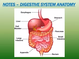
Esophagus • Muscular tube connecting pharynx and stomach • Can be short • Crop- specialization in birds • Outpocketing of esophagus • Used to store food • Pigeon’s milk Figure 11.16: Esophagus and crop of bird.
Stomach • Muscular chamber • Secretes gastric juices • Different lining of stomachs • Esophageal-like epithelia • Glandular epithelia • Ruminant stomach • 4 chambers: rumen, reticulum, omasum, abomasum • Human stomach • Cardiac sphincter- esophagus meets stomach • Mostly lined with gastric epithelium Figure 11.17: Stomach of mammals with esophageal-like epithelia in gray and glandular epithelia in red.
Stomach Structure • Greater and lesser curvature • Messentaries • Greater omentum – attaches along greater curvature • Lesser omentum – attaches along lesser curvature • Cecum- increases surface area • 2 parts in bird and crocodile stomach • Proventiculus-glandular • Gizzard- grinding mill (gastroliths)
4-Chambered Stomachs • Rumen- food enters • Bacterial action • Reticulum- forms a bolus • Omasum- reswallowed grass • Salivary action • Abomasum- food worked out by gastric glands Figure 11.18: Stomach of calf.
Small Intestine • Duodenum- 1st segment • Bile and pancreatic ducts • Jejunum and Ileum subdivisions Figure 11.19: Digestive tract showing regions of small intestine.
Small Intestine • Brunner’s Glands- mucous glands in duodenum and jejunum • Peyer’s Patches- lymphatic nodules in ileum • Crypts of Lieberkühns- intestinal glands at base of villi • Lacteals- within villi—interior lymphatic vessels • Transport fat molecules to circulatory system • Valve of Kirckring- increases surface area
Small Intestine Figure 11.20: Histology of alimentary canal of a mammal showing various glands of small intestine.
Large Intestine • Fish and amphibians - straight and short • Amniotes- divided into colon and rectum • Ileocecal valve- allows passage from small intestine into large • Sigmoid flexure- S-shaped region at rectum • Cecum- aids in absorption • Terminates at vermiform appendix • Cloaca- common chamber for digestive, urinary, and reproductive products to empty (includes monotremes) Figure 11.21: Large intestine of human.
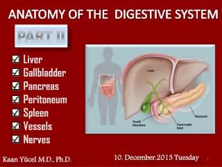
Liver • Liver is diverticulum of primitive gut • Liver produces bile • Bile stored in gallbladder • Common bile duct • Ampulla of Vater- terminal portion Figure 11.22: Development of liver and pancreas.
Pancreas • Pancreas – diverticulum of gut • Duct of Santorini- small, dorsal pancreas • Duct of Wirsung- large, ventral pancreas • Accessory duct- large duct after small, dorsal duct disappears • Exocrine and endocrine glands • Islets of Langerhans- endocrine glands
Literature Cited Figure 11.1, 11.3, 11.4, 11.5, 11.10, 11.15, 11.16, 11.17, 11.18 & 11.22- Kent, George C. and Robert K. Carr. Comparative Anatomy of the Vertebrates. 9th ed. McGraw-Hill, 2001. Figure 11.2- http://www.mouth-cancer-symptoms.com/ Figure 11.6- http://gargravarr.cc.utexas.edu/owl/2002/ Figure 11.7- http://www.proexotics.com/collection_nonPE9.html Figure 11.8- http://www.kingsnake.com/reptilia-italia/My_HomePage_file/snakesgeneral.htm Figure 11.9- http://www.okc.cc.ok.us/biologylabs/Documents/zoology/22 Figure 11.11- http://www2.worldbook.com/features/reptiles/html/body_senorg.html Figure 11.12- http://www.rspb.org.uk/birds/whatyoucando/attracthousemartins/index.asp Figure 11.13- http://people.eku.edu/ritchisong/342notes7.html Figure 11.14- http://www.cortexity.com:8080/nicksblog/ Figure 11.19- http://www.yoursurgery.com/ProcedureDetails.cfm?BR=1&Proc=49 Figure 11.20- Kardong, K. Vertebrates: Comparative Anatomy, Function, Evolution. McGraw Hill, 2002. Figure 11.21- http://www.becomehealthynow.com/popups/lrg_intest.htm