Download
1 / 65
880 likes | 1.48k Vues
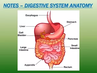
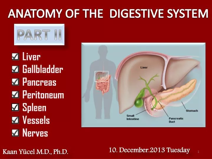
ANATOMY OF THE DIGESTIVE SYSTEM. PART II. Liver Gallbladder Pancreas Peritoneum Spleen Vessels Nerves. 1 0. December . 2013 Tuesday. Kaan Yücel M.D., Ph.D . 1. LIVER. largest gland in the body , second largest organ. A pproximately 1500 g 2.5 % of adult body weight

E N D
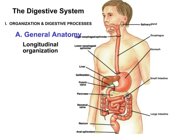
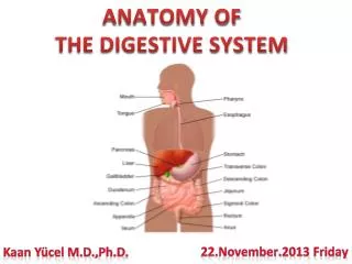
ANATOMY OF THE DIGESTIVE SYSTEM PART II • Liver • Gallbladder • Pancreas • Peritoneum • Spleen • Vessels • Nerves • 10.December.2013Tuesday • Kaan Yücel M.D., Ph.D.
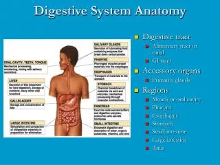
1. LIVER largest gland in the body, secondlargest organ Approximately1500 g 2.5% of adult body weight On the right side Crosses midline toward the left nipple. Righthypochondrium & upper epigastrium. Extends into left hypochondrium.
1. LIVER largest gland in the body, secondlargest organ
1. LIVER Except for fat, all nutrients absorbed from the digestive tract are initially conveyed to the liver by portal venous system.
1. LIVER In addition to its many metabolic activities Stores glycogen. Secretes bile. aids in the emulsification of fat.
Journey of Mr. Bile Bile passes from the liver via the biliary ducts right and left hepatic ducts join to form common hepatic duct unites with cystic duct to form (common) bile duct. Liver produces bile continuously; between meals accumulatedstored concentratedin the gallbladder. When food arrives in the duodenum, gallbladder sends concentrated bile through the biliary ducts to the duodenum.
1. LIVER convex diaphragmatic surfaceanterior, superior, and some posterior relatively flat or even concave visceral surface posteroinferior separated anteriorly by its sharp inferior border that follows the right costal margin.
1. LIVER Visceralsurface covered with visceral peritoneum except in the fossa for the gallbladder at the portahepatis (gateway to the liver). Portahepatis point of entry into the liver for hepatic arteries portal vein, and the exit point for hepaticducts.
ASSOCIATED LIGAMENTS OF THE LIVER FALCIFORM LIGAMENT Liver attached to anterior abdominal wall
ASSOCIATED LIGAMENTS OF THE LIVER Additional folds of peritoneum connect the liverto StomachHepatogastric ligament DuodenumHepatoduodenal ligament DiaphragmRight&left triangular ligaments Anterior&posterior coronary ligaments
Surroundedbyvisceralperitoneum except for a small area of the liver against the diaphragm bare area
1. LIVER divided into right and left lobes by fossae for the gallbladder & inferior vena cava. quadrate and caudate lobes
1. LIVER Quadrate lobe left by fissure for ligamentumteres right by the fossa for the gallbladder. Functionally it is related to the left lobe of the liver. Caudatelobe left by the fissure for ligamentumvenosum right by the groove for the inferior vena cava.
2. GALL BLADDER & THE BILIARY DUCTS a pear-shaped sac lying on the visceral surface of the right lobe of the liver in a fossa between the right and quadrate lobes rounded end fundus of gallbladder major part in the fossabody of gallbladder which may be against transverse colon & superior part of the duodenum narrow part neck of gallbladder
2. GALL BLADDER & THE BILIARY DUCTS In its natural position body of the gallbladder lies anterior to superior part of duodenum its neck and cystic duct are immediately superior to the duodenum.
2. GALL BLADDER & THE BILIARY DUCTS The pear-shaped gallbladder can hold up to 50 mL of bile. The biliary ducts convey bile from the liver to the duodenum. Bile is produced continuously by the liver and stored and concentrated in the gallbladder, which releases it intermittently when fat enters the duodenum. Bile emulsifies the fat so that it can be absorbed in the distal intestine.
2. GALL BLADDER & THE BILIARY DUCTS The bile duct (formerly called the common bile duct) forms by the union of the cystic duct and common hepatic duct. The bile duct descends posterior to the superior part of the duodenum and lies in a groove on the posterior surface of the head of the pancreas.
3. PANCREAS Liesmostly posterior to the stomach Extendsacrosstheposteriorabdominalwall Duodenum on theright, Spleen on theleft
3. PANCREAS produces: Exocrinesecretion (pancreatic juice from the acinar cells) that enters the duodenum through the main and accessory pancreatic ducts. Endocrine secretions (glucagon &insulin from the pancreatic islets [of Langerhans]) that enter the blood.
3. PANCREAS (secondarily) retroperitoneal except for a small part of its tail • Head • Uncinate process • Neck • Body • Tail • The head of pancreas lies within the C-shaped concavity of the duodenum. • Uncinateprocess • Projectsfrom the lower part of the head • passes posterior to the superior mesenteric vessels.
3. PANCREAS Neckof pancreas anterior to superior mesenteric vessels. Posterior to the neck of the pancreas superior mesenteric & splenic veins join to form the portal vein.
3. PANCREAS Tail of pancreas passes between layers of splenorenalligament.
BILIARY DUCTS The main pancreatic duct begins in the tail of the pancreas. passes to the right through the body of the pancreas. Afterentering the head of the pancreas, turns inferiorly. In the lower part of the head of pancreas, joins the bile duct. hepatopancreaticampulla (ampulla of Vater) enters descending (second) part of the duodenum at the major duodenal papilla. Surrounding the ampulla sphincter of ampulla (sphincter of Oddi), a collection of smooth muscle. The accessory pancreatic duct empties into the duodenum just above the major duodenal papilla at the minor duodenal papilla.
4. SPLEEN an ovoid, usually purplish, pulpy mass most vulnerable abdominal organ. located @ superolateralpart of left upper quadrant (LUQ) left hypochondrium enjoys protection of the inferior thoracic cage.
4. SPLEEN connected to greater curvature of the stomach by gastrosplenic ligament contains the short gastric and gastro-omental vessels left kidney by splenorenal ligamentcontains the splenic vessels.
4. SPLEEN surrounded by visceral peritoneum except in the area of the hilum on the medial surface of the spleen. splenic hilum entry point for the splenic vessels and occasionally the tail of the pancreas reaches this area.
4. SPLEEN Largest of the lymphatic organs Participates in the body's defense system as a site of lymphocyte (white blood cell) proliferation and of immune surveillance & response. To accommodate these functions, the spleen is a soft, vascular (sinusoidal) mass with a relatively delicate fibroelastic capsule. The spleen normally contains a large quantity of blood that is expelled periodically into the circulation by the action of the smooth muscle in its capsule and trabeculae.
5. PERITONEUM Abdominal viscera either suspended in the peritoneal cavity by folds of peritoneum (mesenteries) intraperitoneal or outside the peritoneal cavity. Retroperitoneal : onlyonesurfaceorpart o of one surface covered by peritoneum
5. PERITONEUM Parietalperitoneum innervated by somatic afferents carried in branches of the associated spinal nerves and is therefore sensitive to well-localized pain. Visceralperitoneum innervated by visceral afferents that accompany autonomic nerves (sympathetic and parasympathetic) back to the central nervous system.
PERITONEAL CAVITY a potential space of capillary thinness between parietal & visceral layers of peritoneum continues inferiorly into the pelvic cavity. contains a thin film of peritoneal fluid. composed of water, electrolytes, &other substances derived from interstitial fluid in adjacent tissues. lubricates peritoneal surfaces, enabling viscera to move over each other without friction, and allowing the movements of digestion. Largearea Spread of diseases Administeringcertain types of treatment and a number of procedures
PERITONEAL CAVITY Omental bursa continuous with the greater sac through an opening Omental (epiploic) foramen
PERITONEAL CAVITY divided into Greater sac: begins @ diaphragm. Most of theperitonealcavity. Omental bursa (Lessersac): posteriortostomach & liver
PERITONEAL CAVITY Surrounding the omental (epiploic) foramen numerous structures covered with peritoneum. portal vein, hepatic artery proper, and bile duct anteriorly inferior vena cava posteriorly caudate lobe of the liver superiorly first part of the duodenum inferiorly.
GREATER OMENTUM large, apron-like, peritoneal fold Attachesto greater curvature of the stomach &first part of the duodenum. Drapes inferiorly over transverse colon & coils of the jejunum and ileum. Becomesadherent to the peritoneum on the superior surface of the transverse colon & anterior layer of the transverse mesocolon before arriving at the posterior abdominal wall.
MESENTERIES peritoneal folds that attach viscera to the posterior abdominal wall. allow some movement and provide a conduit for vessels, nerves, and lymphatics to reach the viscera Mesentery associated with parts of small intestine Transversemesocolon associated with transverse colon Sigmoidmesocolon associated with sigmoid colon
LESSER OMENTUM from lesser curvature of the stomach & first part of the duodenum toinferior surface of the liver. divided into: medial hepatogastricligament lateral hepatoduodenalligament.
PERITONEAL LIGAMENTS consist of two layers of peritoneum connect two organs to each other or attach an organ to the body wall may form part of an omentum. usually named after the structures being connected. For example, splenorenalligament connects the left kidney to the spleen and gastrophrenicligament connects the stomach to the diaphragm.
6. PORTAL SYSTEM Portalvein final common pathway for the transport of venous blood from the spleen, pancreas, gallbladder, and the abdominal part of the gastrointestinal tract. formed by union of splenic vein & superior mesenteric vein posterior to the neck of the pancreas.
7. VESSELS & NERVES OF THE GASTROINTESTINAL SYSTEM Arterial blood is supplied mainly by the coeliac artery to the stomach, pancreas, spleen and liver and by the mesenteric arteries to the intestines. Abdominal aorta
7. VESSELS & NERVES OF THE GASTROINTESTINAL SYSTEM Arterial blood is supplied mainly by the coeliac artery to the stomach, pancreas, spleen and liver and by the mesenteric arteries to the intestines.
7. VESSELS & NERVES OF THE GASTROINTESTINAL SYSTEM The duodenum is supplied by branches of the superior mesenteric artery and those of the coeliac trunk. The jejunum and ileum are supplied by the branches of superior mesenteric artery.
7. VESSELS & NERVES OF THE GASTROINTESTINAL SYSTEM Ascendingcolon several branches from the superior mesenteric artery such as right colic artery, ileocolic artery, etc. Transverse colon branches from the superior mesenteric artery and left colic artery; a branch of the inferior mesenteric artery. descending colon. Sigmoidalarteries from inferior mesenteric artery.
7. VESSELS & NERVES OF THE GASTROINTESTINAL SYSTEM The arterial supply to the rectum and anal canal from superior to inferior branches from inferior mesenteric artery internal iliac artery internal pudendal artery (a branch of the internal iliac artery).
7. VESSELS & NERVES OF THE GASTROINTESTINAL SYSTEM Venous blood drains from the stomach, pancreas and spleen via the hepatic portal vein into the liver, where the products of digestion undergo further processing and detoxification. Blood from the oesophagus and rectum (middle & inferiorparts)does not go through the liver but drains directly into the venous system.
7. VESSELS & NERVES OF THE GASTROINTESTINAL SYSTEM superior mesenteric vein drains blood from small intestine, cecum, ascending colon, and transverse colon inferiormesenteric vein drains blood from rectum, sigmoid colon, descending colon, and splenic flexure
7. VESSELS & NERVES OF THE GASTROINTESTINAL SYSTEM inferiormesenteric vein begins as the superior rectal vein and ascends, receiving tributaries from the sigmoid veins and the left colic vein Inf. & middlerectalveins Internaliliacvein
Foregut: anteriorpart of thealimentarycanal, frommouthtoduodenum @ entrance of the bile duct. Midgut: fromdistalhalf of 2ndpart of duoedenum, toproximaltwo-thirds of thetransversecolon. Hindgut: distalthird of thetransversecolonandthesplenicflexure, thedescendingcolon, sigmoid colon, andrectum.