Download
1 / 15
150 likes | 185 Vues
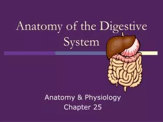
Chapter 25 Anatomy of the Digestive System. Overview of the Digestive System. Role of the digestive system Prepares food for absorption and use by all the cells of the body Food material not absorbed becomes feces that is eliminated

E N D
Overview of the Digestive System • Role of the digestive system • Prepares food for absorption and use by all the cells of the body • Food material not absorbed becomes feces that is eliminated • Digestion depends on both endocrine and exocrine secretions and the controlled movement of ingested food materials through the gastrointestinal (GI) tract
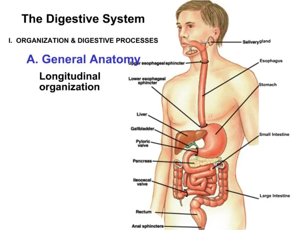
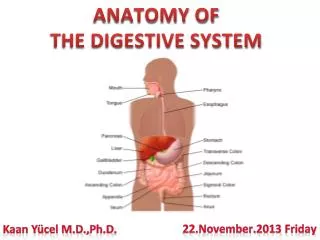
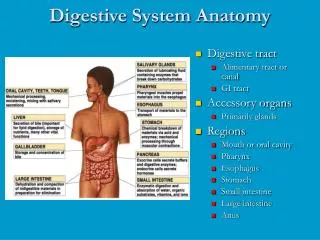
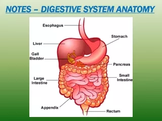
Overview of the Digestive System • Organization of the digestive system • Organs of digestion (Figure 25-1, Table 25-1) • Form the alimentary canal or gastrointestian (GI) tract • Extends through the abdominopelvic cavity • Ingested food material passing through the lumen of the GI tract is outside the internal environment of the body
Overview of the Digestive System • Organization of the digestive system • Wall of the GI tract (Figure 25-2, Table 25-2) • Layers • Mucosa • Mucous epithelium – epithelial tissue • Lamina propria – connective tissue • Muscularis mucosae – muscle tissue • Submucosa • Connective tissue with glands, vessels, and nerves • Muscularis • Smooth muscle • Myenteric plexus • Serosa • Connective tissue and peritoneum • Visceral layer of peritoneum
Mouth • Structure of the oral cavity (Figure 25-3) • Lips • Covered externally by skin and internally by mucous membrane • Junction between skin and mucous membrane is highly sensitive • Philtrum and tubercle • When lips are closed, line of contact is oral fissure • Cheeks • Lateral boundaries of oral cavity • Continuous with lips and lined by mucous membrane • Formed in large part by buccinator muscle covered by adipose tissue • Contain mucus-secreting glands
Mouth • Structure of the oral cavity • Hard and soft palates • Hard palate consists of portions of two maxillae and two palatines • Soft palate is made of muscle arranged in an arch • Fauces and uvula • Tongue • Solid mass of skeletal muscle covered by a mucous membrane (Figure 25-4) • Important for mastication and deglutition • Root, tip, and body • Papillae located on dorsal surface of tongue • Lingual frenulum anchors tongue to floor of mouth
Mouth • Salivary glands • Three pairs of compound tubuloalveolar glands (Figure 25-6) secrete approximately 1 liter of saliva each day • Buccal glands contribute less than 5% of total salivary volume but provide for hygiene and comfort of oral tissues • Parotid glands • Largest of the paired salivary glands • Produce watery saliva containing enzymes (no mucus) • Submandibular glands • Compound glands that contain enzyme and mucus-producing elements • Sublingual glands • Smallest of the salivary glands • Produce a mucous type of saliva (no enzymes)
Mouth • Teeth • Typical tooth (Figure 25-7) • Crown • Exposed portion of a tooth • Covered by enamel • Neck • Narrow portion that joins the crown to the root • Surrounded by the gingivae (gums) • Root • Fits into socket of alveolar process • Suspended by fibrous periodontal membrane • Dentin and Cementum • Pulp cavity • Located in dentin • Contains connective tissue, blood and lymphatic vessels, and sensory nerves
Mouth • Types of teeth (Figure 25-8) • Deciduous teeth • 20 baby teeth, which appear early in life • First teeth appear at about 6 months of age • One each month after that • Shed between the ages of 6 and 13 years • Permanent teeth • 32 teeth, which replace the deciduous teeth • Central incisors • Lateral incisors • Canines • Premolars (1st, 2nd) • Molars (1st,2nd,3rd)