Download
1 / 35
350 likes | 371 Vues
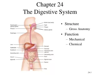
The Digestive System Chapter 25. Function of the Digestive System. To break down food into a “usable” (absorbable) form To supply our cells with the nutrients they need for energy, growth & repair. Organs of the Digestive System.

E N D
Function of the Digestive System • To break down food into a “usable” (absorbable) form • To supply our cells with the nutrients they need for energy, growth & repair
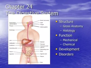
Organs of the Digestive System • Gastrointestinal tract (GIT) – continuous passageway which contains the food from the time it enters the body, until it leaves; organs include: • mouth (oral cavity), pharynx, esophagus, stomach, small intestine, large intestine, rectum, anus • Accessory organs - participate in digestive processes; organs include: • teeth, tongue, salivary glands, liver, gall bladder, pancreas
Processes of Digestion • Ingestion • Movement along GIT • Voluntary – e.g. swallowing • Involuntary – e.g. peristalsis • 3. Secretion – release of water, enzymes, acids, buffers, mucous, etc. into GIT for physical (mechanical) & chemical digestive processes
Processes of Digestion • 4. Digestion • Mechanical processing – physical breakdown of food; e.g. mastication, emulsification, mixing waves, segmentation • Chemical digestion – chemical breakdown of food; disassembling of organic molecules into their component parts; requires enzymes • carbohydrates disaccharides monosaccharides • proteins amino acids • lipids fatty acids & monoglycerides
Processes of Digestion 5. Absorption – movement of nutrients from GIT into blood capillaries (monosaccharides, amino acids, H2O, vitamins, minerals) or lymphatic capillaries (fatty acids) 6. Excretion (Defecation) – removal of waste products from GIT
epithelium – stratified squamous or simple columnar • lamina propria – loose CT • muscuaris mucosa – smooth muscle Mucosa Submucosa CT with BV’s, nerves & lymphatics Muscularis externa Skeletal muscle at beginning & end of GIT, smooth muscle (inner circular; outer longitudinal layer) from lower esophagus to rectum Serosa (a.k.a. viseral peritoneum) Histology of the GIT 4 layers of tissue surround the lumen of the GIT
Peritoneum & Mesenteries • The abdominal cavity is lined with parietal peritoneum & many of the organs within are covered with visceral peritoneum • Folds of peritoneum called “mesenteries” attach some organs to others • greater omentum • lesser omentum • mesentery proper • mesocolon
Mouth (oral cavity) • Regions include the vestibule & oral cavity proper • Roof comprised of hard & soft palate; floor primarily comprised of tongue • Mucosa of stratified squamous epithelium (non-keratinized) • Joins to the oropharynx at the fauces
Tongue – • stratified sqamous epith. over skeletal muscle • intrinsic & extrinsic muscles • papillae • filiform • fungiform • circumvallate
Salivary glands – secrete saliva – made of H2O, salts & “salivary amylase” Parotid gland Parotid duct Sublingual gland Submandibular duct Submandibular gland
Teeth – involved in “mastication” • 2 sets of teeth – deciduous & permanent • 4 types of teeth – incisors, cuspids (canines), bicuspids (premolars), molars
Parts of a tooth – • crown – dentin surrounded by enamel, has hollowed pulp cavity filled with CT pulp • neck – at gingival border • root – within mandible & maxilla, has hollowed root canal with BVs & nerves, root canal opens at apical foramen
Pharynx • Common passageway for air & food • oropharynx & laryngopharynx lined with stratified squamous epithelium (nasopharynx lined with PSCC) • uvula & epiglottis protect airway when swallowing (“deglutition”) nasopharynx uvula oropharynx epiglottis laryngopharynx
Esophagus • muscular tube running from pharynx, posterior to trachea, down thoracic cavity, through “esophageal hiatus” of diaphragm, to lower esophageal (a.k.a. cardiac) sphincter at junction of stomach • functions in “deglutition” through action of peristalsis • mucosa is stratified squamous epithelium • variations in muscularis externa – begins as skeletal muscle at upper 1/3, mixed skeletal & smooth muscle in middle, smooth muscle by lower 1/3
Stomach - Gross Anatomy Lower esophageal (cardiac) sphincter Pyloric sphincter
Stomach - Histology Rugae – folds of mucosa & submucosa to allow for expansion of stomach Mucosa of simple columnar epithelium with mucous cells Gastric pit leading to gastric glands
Stomach – Histology (cont) - Secrete mucus to protect epithelial cells from enzymes & acid - Secrete HCl (for protein digestion) & intrinsic factor (for B12 absorption) - Secrete pepsinogen which gets converted to “pepsin” when mixed with HCl; for protein digestion (peptic cells) Entero- - Secrete gastrin to regulate stomach emptying (G-cells)
pepsin proteins polypeptides HCl Stomach • Modifications in stomach include 3 layers of smooth muscle in muscularis externa – outer longitudinal, middle circular, innermost oblique layer • Functions of stomach include • temporary storage of food • mechanical breakdown of food to “chyme” through powerful mixing waves • intrinsic factor for vitamin B12 absorption • start of chemical digestion of proteins –
Small Intestine - Anatomy • connects stomach to large intestine; 15-20’ long; 1” diameter; held together in abdominal cavity by “mesentery proper” • site for completion of chemical digestion & absorption of nutrients • comprised of three regions: Duodenum – 10” in length; receives chyme from stomach, secretions from liver, gallbladder & pancreas Jejunum – 8’ long; most digestion & absorption occurs here Ileum – 12’ long; connects to cecum of large intestine at iliocecal valve (sphincter)
Small Intestine Modifications in mucosa & submucosa of intestinal wall designed to increase functional surface area: • Plicae circulares (circular folds) – large transverse ridges; most abundant in jejunum • Villi – small finger-like projections of mucosal folds across surface of intestine Plicae circulares
Small Intestine Villi • Villi lined with “absorptivecells” - mucosal epithelium of simple columnar epithelium with microvilli “brush border” . These cells also produce enzymes (disaccharidases & peptidases) for final digestion of carbs and proteins • Submucosa of each villus contains a capillary network & a “lacteal” (lymphatic capillary) for absorption of nutrients
Intestinal crypts containing stem cells and intestinal glands • Between villi are intestinal crypts. Stem cells here can replace old cells found lining villi • Intestinal glands within intestinal crypts secrete “intestinal juice” – provides watery medium to keep enzymes & digestive products in solution for help with absorption. Small Intestine Villi
Stomach Tail Body Head • Retroperitoneal elongated organ lying posterior to stomach, from duodenum to spleen • Both endocrine (pancreatic islets of Langerhans – secretes insulin & glucagon) & exocrine gland (pancreatic acini – secretes pancreatic juice (aka pancreatin) through pancreatic duct(s) to duodenum Pancreas Pancreatic duct Duodenum
Pancreas • Pancreatic juice – mixture of enzymes & buffers (sodium bicarbonate) secreted by acinar cells into pancreatic duct & released into duodenum • pancreatic amylase Starch maltose • lipase Lipidsfatty acids + monoglycerol • proteases (trypsin, chymotrypsin, carboxypeptidase) Proteins & polypeptides small peptides tri & dipeptides • nucleases – digest RNA & DNA • sodium bicarbonate – neutralizes acidic chyme because enzymes in small intestine need an alkaline pH
Liver - Anatomy • Largest organ within the body • Comprised of 4 lobes: • Large right & left lobes divided by falciform ligament; small caudate (by IVC) & quadrate (by gall bladder ) lobes • falciform ligament continues at inferior margin as ligamentum teres (round ligament) (remnant of umbilical vein) • Lobes of liver functionally divided into microscopic lobules
Liver - Histology • Lobules comprised of rows of Hepatocytes arranged radially around a central vein • Hepatocytes surround blood sinusoids (capillary structures) which are partially lined with phagocytic Kupffer (aka stellate reticuloendothelial) cells hepatocytes central vein sinusoids
Liver • One function of hepatocytes is to produce bile, which gets secreted into bile canaliculi of lobule • Bile canaliculi merge to form bile ducts, which are part of the portal triad seen at each corner of the lobules. Bile ducts merge to eventuallycreate the right & left hepatic ducts
Liver & gall bladder • Right & left hepatic ducts unite to form common hepatic duct which merges with cystic duct of gall bladder to form common bile duct which joins with pancreatic duct & enters the duodenum Right hepatic duct Left hepatic duct • Gall bladder – hollow muscular sac under right lobe of liver; stores & concentrates bile; releases bile through cystic duct • Bile released into duodenum functions in emulsification of lipids, absorption of fats (due to presence of bile salts), & excretion of bilirubin
Stomach Tail Body Pancreatic & bile ducts Common bile duct Accessory pancreatic duct Head Pancreatic duct
Liver - Functions • The liver has over 200 functions including (but not limited to): • Bile production & excretion • Metabolic regulation – • storage of glycogen, fatty acids, fat-soluble vitamins & minerals • interconversion of nutrients (“gluconeogenesis”) • detoxification & removal of drugs, toxins & hormones • hematological regulation – • phagocytosis of worn-out RBCs, bacteria & other pathogens • synthesis of plasma proteins
Blood Supply to Liver • In order for the liver to perform all of its functions, it receives blood through 2 vessels: • Hepatic artery - delivers oxygenated blood into sinusoids of liver • Hepatic Portal vein – delivers de-oxygenated, nutrient-rich blood from digestive organs to sinusoids of liver Liver uses O2 & nutrients within blood of sinusoids & then blood drains into central veins of lobule which merge to form the hepatic veins, which drain into the IVC
Transverse colon Ascending colon Descending colon ileum Ileocecal sphincter Cecum Sigmoid colon Vermiform appendix Large Intestine • 3 regions: cecum- blind pouch; has appendix attached • colon – ascending, (hepatic flexure), transverse, (splenic flexure), descending, sigmoid • rectum – last 1” known as “anal canal” • Begins at the ilium & ends at the anus; 5’ long; 3” in diameter Hepatic (rt. Colic) flexure Splenic (lt. colic) flexure Rectum Anal canal Internal anal sphincter Rectum Rectum Anal canal External anal sphincter Anus
Large Intestine • main functions – H2O re-absorption; absorption of some vitamins & minerals; formation & temporary storage of fecal material • no chemical (enzymatic) digestion but some bacterial • Simple columnar epithelium in mucosa, except at anal canal (strat. Squam.) • No plicae circularis or villi • Modifications in muscularis externa & serosa : • longitudinal muscle layer forms bands called “taeniae coli” which create puckers known as “haustra” • serosa forms “epiploic appendages” haustra taeniae coli epiploic appendages THE END (literally!)