Download
1 / 89
930 likes | 1.19k Vues
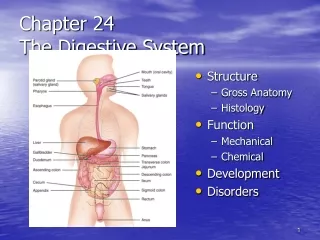
Chapter 25 The Digestive System. General anatomy & digestive processes Mouth through esophagus Stomach Liver, gallbladder & pancreas Small intestine Chemical digestion & absorption Large intestine. Digestive Functions. Ingestion = intake of food Digestion = breakdown of molecules

E N D
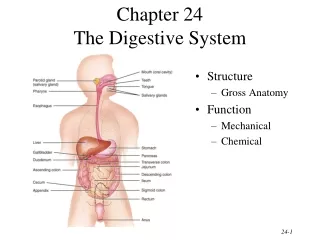
Chapter 25The Digestive System • General anatomy & digestive processes • Mouth through esophagus • Stomach • Liver, gallbladder & pancreas • Small intestine • Chemical digestion & absorption • Large intestine
Digestive Functions • Ingestion = intake of food • Digestion = breakdown of molecules • Absorption = uptake nutrients into blood/lymph • Defecation = elimination of undigested material
Stages of Digestion • Mechanical digestion is physical breakdown of food into smaller particles • teeth & churning action of stomach & intestines • Chemical digestion is series of hydrolysis reactions that break macromolecules into their monomers • enzymes from saliva, stomach, pancreas & intestines • results • polysaccharides into monosaccharides • proteins into amino acids • fats into glycerol and fatty acids
Digestive Processes • Motility = muscular contractions that break up food, mix it with enzymes & move it along • Secretion = digestive enzymes & hormones • Membrane transport = absorption of nutrients
Subdivisions of the Digestive System • Digestive tract (GI tract) • 30 foot long tube extending from mouth to anus • Accessory organs • teeth, tongue, liver, gallbladder, pancreas, salivary glands
Tissue Layers of the GI Tract • Mucosa • epithelium • lamina propria • muscularis mucosae • Submucosa • Muscularis externa • inner circular layer • outer longitudinal layer • Adventitia or Serosa • areolar tissue or mesothelium
Relationship to the Peritoneum • Only duodenum, pancreas & parts of large intestine are retroperitoneal • Dorsal mesentery suspends GI tract & forms serosa (visceral peritoneum) of stomach & intestines • Ventral mesentery forms lesser & greater omentum • lacy layer of connective tissue contains lymph nodes, lymphatic vessels and blood vessels
Lesser & Greater Omentum • Lesser attaches stomach to liver • Greater covers small intestines like an apron
Mesentery and Mesocolon • Mesentery of small intestines holds many blood vessels • Mesocolon anchors the colon to the back body wall
Regulation of Digestive Tract • Neural control • short myenteric reflexes (swallowing) • long vagovagal reflexes (parasympathetic stimulation of digestive motility and secretion) • Hormones • messengers diffuse into bloodstream, distant targets • Paracrine secretions • messengers diffuse to nearby target cells
Permanent & Baby Teeth • Baby teeth (20) by 2 years; Adult (32) between 6 and 25 • Occlusal surfaces and cusp numbers differ
Tooth Structure • Periodontal ligament is modified periosteum • anchors into alveolus • Cementum & dentin are living tissue • Enamel is noncellular secretion formed during development • Root canal leads into pulp cavity • nerves & blood vessels • Gingiva or gums
Mastication or Chewing • Breaks food into smaller pieces to be swallowed • surface area exposed to digestive enzymes • Contact of food with sensory receptors triggers chewing reflex • tongue, buccinator & orbicularis oris manipulate food • masseter & temporalis elevate the teeth to crush food • medial & lateral pterygoids swing teeth in side-to-side grinding action of molars
Saliva • Functions of saliva • moisten, begin starch & fat digestion, cleanse teeth, inhibit bacteria, bind food together into bolus • Hypotonic solutions of 99.5% water and solutes: • amylase = begins starch digestion • lingual lipase = digests fat after reaches the stomach • mucus = aids in swallowing • lysozyme = enzyme that kills bacteria • immunoglobulin A = inhibits bacterial growth • electrolytes = Na+, K+, Cl-, phosphate & bicarbonate • pH of 6.8 to 7.0
Salivary Glands • Small intrinsic glands found under mucous membrane of mouth, lips, cheeks and tongue -- secrete at constant rate • 3 pairs extrinsic glands connected to oral cavity by ducts • parotid, submandibular and sublingual
Histology of Salivary Glands • Compound tubuloacinar glands • Mucous cells secrete mucus • Serous cells secrete thin fluid rich in amylase • Mixed acinus is possible
Salivation • Total of 1 to 1.5 L of saliva per day • Cells filter water from blood & add other substances • Food stimulates receptors that signal salivatory nuclei in the medulla & pons • parasympathetic stimulation salivary glands produce thin saliva, rich in enzymes • sympathetic stimulation produce less abundant, thicker saliva, with more mucus • Higher brain centers stimulate salivatory nuclei so sight, smell & thought of food cause salivation
Pharynx • Skeletal muscle • deep layer – longitudinal orientation • superficial layer – circular orientation • superior, middle and inferior pharyngeal constrictors
The Esophagus • Straight muscular tube 25-30 cm long • nonkeratinized stratified squamous epithelium • esophageal glands in submucosa • skeletal muscle in upper part & smooth in bottom • Extends from pharynx to cardiac stomach passing through esophageal hiatus in the diaphragm • inferior pharyngeal constrictor excludes air from it • Lower esophageal sphincter closes orifice to reflux
Swallowing or Deglutition • Series of muscular contractions coordinated by swallowing center in medulla & pons • motor signals from cranial nerves V, VII, IX and XII • Buccal phase • tongue collects food & pushes it back into oropharynx • Pharyngeal-esophageal phase • soft palate rises & blocks nasopharynx • infrahyoid muscles lift larynx & epiglottis is folded back • pharyngeal constrictors push bolus down esophagus • liquids in 2 seconds -- food bolus may take 8 seconds • lower esophageal sphincter relaxes
Introduction to the Stomach • Mechanically breaks up food particles, liquifies the food & begins chemical digestion of protein & fat • resulting soupy mixture is called chyme • Stomach does not absorb any significant amount of nutrients • does absorb aspirin & some lipid-soluble drugs
Gross Anatomy of the Stomach • Muscular sac (internal volume from 50ml to 4L, FULL) • J - shaped organ with lesser & greater curvatures • regional differences • cardiac region just inside cardiac orifice • fundus is domed portion superior to esophageal opening • body is main portion of organ • pyloric region is narrow inferior end • antrum & pyloric canal • Pylorus is opening to duodenum • thick ring of smooth muscle forms a sphincter
Innervation and Circulation • Innervation by parasympathetic fibers from vagus & sympathetic fibers from the celiac plexus • All blood drained from stomach is filtered through the liver before returning to heart
Gross Anatomy of Stomach • Notice: bulge of fundus, narrowing of pyloric region, thickness of pyloric sphincter and greater & lesser curvatures
Unique Features of Stomach Wall • Mucosa • simple columnar glandular epithelium • lamina propria is filled with tubular glands (gastric pits) • Muscularis externa has 3 layers • outer longitudinal, middle circular & inner oblique layers
Cells of the Gastric Glands • Mucous cells secrete mucus • Regenerative cells divide rapidly to produce new cells that migrate upwards towards surface • Parietal cells secrete HCl acid & intrinsic factor • Chief cells secrete chymosin & lipase in infancy & pepsinogen throughout life • Enteroendocrine cells secrete hormones & paracrine messengers
Gastric Secretions • 2 to 3 L of gastric juice/day (H2O, HCl & pepsin) • Parietal cells contain carbonic anhydrase (CAH) CAH • CO2 + H2O H2CO3 HCO3- + H+ • H+ is pumped into stomach lumen by H+K+ATPase • antiporter uses ATP to pump H+ out & K+in • HCO3- exchanged for Cl-(chloride shift) • Cl- pumped out to join H+ forming HCl • HCO3- in blood causes alkaline tide (blood pH )
Functions of Hydrochloric Acid • Activates enzymes pepsin & lingual lipase • Breaks up connective tissues & plant cell walls • liquifying food to form chyme • Converts ingested ferric ions (Fe+3) to ferrous ions (Fe+2) that can be absorbed & utilized for hemoglobin synthesis • Destroys ingested bacteria & pathogens
Gastric Enzymes & Intrinsic Factor • Intrinsic factor (parietal cells ) • essential for absorption of B12 by small intestine • necessary for RBC production (pernicious anemia) • Pepsin --- (chief cell) protein digestion • secreted as pepsinogen (an inactive zymogen) • HCl converts it to pepsin (active form) • pepsin then activates more pepsinogen • Gastric lipase & chymosin (chief cell) • lipase digests butterfat of milk in infant • chymosin curdles milk by coagulating its proteins
Chemical Messengers • Many produced by enteroendocrine cells • hormones enter blood distant cells • paracrine secretions neighboring cells • Gut-brain peptides • signaling molecules produced in digestive tract + CNS
Gastric Motility • Swallowing center signals stomach to relax • Arriving food stretches the stomach activating a receptive-relaxation response • resists stretching briefly, but relaxes to hold more food • Rhythm of peristalsis controlled by pacemaker cells in longitudinal muscle layer • gentle ripple of contraction every 20 seconds churns & mixes food with gastric juice • stronger as reaches pyloric region squirting out 3 mL • duodenum neutralizes acids and digests nutrients little at time • typical meal is emptied from stomach in 4 hours
Vomiting • Induced by excessive stretching of stomach, psychological stimuli or chemical irritants (bacterial toxins) • Emetic center in medulla causes lower esophageal sphincter to relax as diaphragm & abdominal muscles contract • contents forced up the esophagus • may even expel contents of small intestine
Regulation of Gastric Function (Phases 1-2) • Cephalic phase • vagus nerve stimulates gastric secretion & motility just with sight, smell, taste or thought of food • Gastric phase • activated by presence of food or semidigested protein • by stretch or in pH • secretion is stimulated by • ACh (from parasympathetic fibers), histamine (from gastric enteroendocrine cells) and gastrin (from pyloric G cells) • receptors for each substance on parietal cells & chief cells
Regulation of Gastric Function (Phase 3) • Intestinal phase - duodenum regulates gastric activity through hormones & nervous reflexes • at first gastric activity increases(if duodenum is stretched or amino acids in chyme cause gastrin release) • enterogastric reflex = duodenum inhibiting stomach • caused by acid and semidigested fats in duodenum • chyme stimulates duodenal cells to release secretin, cholecystokinin (CCK) & gastric inhibitory peptide • all 3 suppress gastric secretion & motility
Liver, Gallbladder and Pancreas • All release important secretions into small intestine to continue digestion