Download
1 / 48
550 likes | 906 Vues
Structure and Function of Human Skin. Lianjun Chen Huashan Hospital. Importance of the study of skin diseases. Common and prevalent diseases Skin beauty and health care reflection of the body health ( paraneoplastic skin diseases)

E N D
Structure and Function of Human Skin Lianjun Chen Huashan Hospital
Importance of the study of skin diseases • Common and prevalent diseases • Skin beauty and health care • reflection of the body health (paraneoplastic skin diseases) • Sex Transmitted Disease(STD), AIDS, other contagious diseases
OUTLINE • Introduction of the Skin • Microstructure of the skin • Epidermis • The basement membrane zone • Dermis • Subcutis • Appendageal Structure • The function of the skin
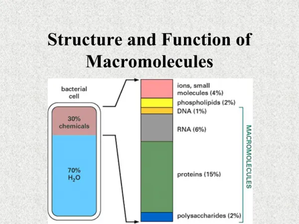
Introduction • The single biggest organ in the body • Cover an average area of 1.5 m2 • Varying thickness (0.4-4mm) • Different color by the race, age, gender & location • Perform many vital roles
Microstructure of the skin • Epidermis • Dermis • Subcutis • The basement membrane zone • Appendageal Structure
Epidermis • Derived from ectoderm • The celluar external layer • Keratinocyte • non-keratinocyte • Melanocyte • Langerhans’ cell • Merkel cell
Keratinocytes • stratum basalum, basal cell layer • stratum spinosum, prickle cell layer • stratum granulosum, granular layer • stratum lucidum • stratum corneum, horny layer
keratinocyte non-keratinocyte keratin + ─ tonofilament + ─ desmosome+ ─ dendritic-shaped ─+
Function of the keratinocytes • Continuously producing keratin and forming horny layer to protect against various chemical and physical factors • Participating immune response in the skin • Secreting cytokines and helping to activate T lymphocytes
Desmosome • Attachment plates between adjacent keratinocytes in basal layer and prickle layer • Desmosome transmembrane protein • desmoglein,DG • desmocollin,DC • Desmosome cytoplasm plakin • desmoplakin,DP • plakoglobulin,PG
Non-keratinocytes • Melanocyte • Langerhans cell • Merkel cell
Melanocytes • Epidermal melanin unit • melanocytes are derived from the nerual crest and reside in the basal layer • Melanocytes are dendritic cells and contain large number of melanosomes • Any one melanocyte is contact with 30-40 keratinocytes through its dendrites, form the so-called epidermal melanin unit. • Protective barrier against UV
Langerhans’ cells • Langerhans’ cells are originate in bone marrow. They exist in the epidermis above the basal layer . • They are characterized by a folded nucleus and distinct intracytoplasmic organelles called Birbeck granules,resembling a tennis racquet. • Recognition, uptake, processing, and presentation of antigens to sensitized T lymphocytes.
Langerhans’ Granules
Merkel cells • a kind of neurosecretory cell • located directly above the basement membrane and closely associated with neurites, so called Merkel cell-axon complex.
Basement membrane zone • The base of basal cell containing hemidesmosome • The lamina lucida • A clear zone crossed by a fine network of anchoring filaments, largely composed of the structural protein laminin • The lamina densa • An electron dense band, largely composed of type IV collagen • The reticular lamina(collagen VII)
Dermis • Derived from mesoderm • Papillary dermis & reticular dermis • Connective tissue • Collagen, elastic fiber, reticulum fiber • matrix (mucopolysaccharides) • blood vessel, lymphatics, nerve ending • Appendageal sructure • apocrine gland, eccrine gland, sebaceous gland, hair follicle, nail, et al
Subcutis • It consist largely of fat tranversed by nerves and blood vessels. • The name implies that it is not properly the part of the skin, however, it could be an integrated whole as for the function.
Appendages • Hair follicles • Sebaceous glands • Sweat glands • Eccine glands • Apocrine glands • Nails Pilosebaceous units
Hair follicles • Classification • Terminal hair Scalp,axillae, pubic,beard eyebrow,eyelash,vibrissa • Vellus • lanugo
Hair Anatomy • Infundibulum – surface to entrance of sebaceous duct • Isthmus – sebaceous duct to pilar muscle insertion • Lower segment – Stem,Bulb (matrix and papilla) • Perifollicular sheath (mesenchyme)
Infundibulum Isthmus Arrector pili muscle Stem Bulb
anagen catagen telogen
Sebaceous glands • Distribute throughout all skin sites except palms and soles,greatest abundant on scalp, face,middle part of the body and vulvae. • Always associated with upper portion of the hair follicles • Reach skin surface,such as • vermilion border of the lip(Fordyce’s spots) • areolas(Montgomery’s tubercles) • prepuce(Tyson’s glands) • eyelids(Meibomian’s glands) • Production of sebum
Eccrine glands • Distribution: almost all skin site • Exclude the vermilion border of the lip, nympha, phallus, glans, flexion side of prepuce and nail beds • most abundant on the palms, soles and axillae • Reach to skin surface directly • Under psychological and thermal control
Eccrine glands • Composition of the eccrine gland • Coil gland is composed of a single layer of cells with two types: pale cell and darker-staining cell and regulated by cholinergic nerve • The duct is composed of a double layer of basophilic epithelial cells and divided into intraepidermal component and intradermal component
Function of the eccrine gland • Dissipate excessive body heat • Soften horniness • Acidify sweat • Take place of partial renal function • Emulsify lipid • Secrete immunoglobulin • : • ⑴
Apocrine glands • Distribution: generally confined to axillae, areolae and the anogenital region. • reach to the hair follicle instead of skin surface • Becoming active at puberty • Producing a protein-rich secretion , sometimes gives out a characteristic odour.
Nails • Consist of a dense plate of hardened keratin between 0.3 and 0.5mm thick • The nail is made up of a nail bed, nail matrix and a nail plate. • The thickened epidermis which underlies the free margin of the nail at the proximal end is called the hyponychium. • Fingernails grow at 0.1 mm per day; the toenails more slowly.
Skin is the largest organ in the body by weight and surface area. • Skin is composed by three layers : epidermis , dermis and subcutis. • Epidermis is the outer layer composed mainly of keratinocytes but also melannocytes and Langerhan’s cells. • Dermis is a supportive connective tissue layer comprised mainly of collogen but also containing elastin embbed in a matrix of mucopolysaccharides.
Hairs are ketatinized tubes emerging from invaginations of the epidermis called follicles. • Sebaceous glands deliver sebum into follicle lubricating the hair surface. • Eccrine sweat glands are found almost over the entire skin surface and innervated by the sympathetic nervous system,responding to temperature/psychological changes. • Apocrine sweat glands are localized (axillae, nipples,perineum,scalp) and androgen-sensitive. • Nails, like hair, are made largely of keratin.
Function of the skin • As a barrier • Provides a protective barrier to: • mechanical,thermal,and other physical injury • the ingress of noxious agents • the excessive loss of moisture and protein(if the skin absent or seriously compromised) • The harmful effect of ionizing radiation especially ultraviolet radiatio • Sensation • Rich in nerve ending and specialized sensory receptor
Function of the skin • Thermoregulation • Skin is the principle organ of heat control • Immunological surveillance • Contains immunologically component cells • Biochemical reaction • Vitamin D synthesis • Involved in androgen degradation and others • Social and sexcial communication • Disorders of the skin such as dicoloration may cause tremendous stress of human, impair the individual’s sense of well-being and self-worth.
Some pictures are from Mosby’s Color Atlas and Textbook of Dermatology. These cannot be copied without the pemission of the publisher, only used in self-learning. • Web address of this curriculum http://jpkc.fudan.edu.cn/s/283/main.jspy • From 12th .Oct, practice time • Place of Assembly: dermatopathology Unit, floor 3, building 6, Huashan Hospital • Time of Assembly: 1:30 p.m. • Tel: 13817311071 Lianjun Chen
Thanks for your attention!