Download
1 / 24
420 likes | 1.03k Vues
Development of Respiratory System. Dr. Sama ul Haque. Objectives. Discuss the formation of the lung buds. Describe the development of larynx. Explain the mechanism of formation of trachea, bronchi and lungs. Differentiate between the periods of lung maturation.

E N D
Development of Respiratory System Dr. Sama ul Haque
Objectives • Discuss the formation of the lung buds. • Describe the development of larynx. • Explain the mechanism of formation of trachea, bronchi and lungs. • Differentiate between the periods of lung maturation. • Discuss the congenital anomalies of the respiratory system.
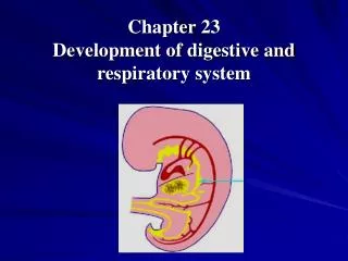
Formation of the Lung Buds • In 4 weeks old embryo, the respiratorydiverticulum (lung bud) appears as an outgrowth from the ventral wall of the foregut. • Epitheliumof the internal lining of the larynx, trachea, and bronchi, as well as that of the lungs, is entirely of endodermal origin.
Formation of the Lung Buds • The cartilaginous, muscular, andconnective tissuecomponents of the trachea and lungs are derived from splanchnic mesoderm surrounding the foregut. • The tracheoesophageal septum, divides the foregut into a dorsal portion, the esophagus, and a ventral portion, the trachea and lung buds.
Development of Larynx • The internal lining of the larynx: From Endoderm • The cartilages and muscles: From mesenchyme of the 4th and 6th Pharyngeal arches. • Proliferating mesenchyme of the two arches transforms into the thyroid, cricoid, and arytenoid cartilages. • Temporary occlusion of the laryngeal lumen occurs due to the proliferation of laryngeal epithelium.
Development of Larynx • Recanalization produce a pair of lateral recesses, the laryngeal ventricles (recesses are bounded by folds of tissue that differentiate into the false and true vocal cords). • Since musculature of the larynx is derived from mesenchyme of the 4th & 6th pharyngeal arches, all laryngeal muscles are innervated by branches of the 10th cranial nerve (vagus nerve). The superior laryngealnerve innervates derivatives of the fourth pharyngeal arch, and the recurrent laryngeal nerveinnervates derivatives of the sixth pharyngeal arch.
Development of Larynx Pharyngeal arches: Each arch contains a cartilaginous component, a cranial nerve, an artery, and a muscular component.
Development of Trachea, Bronchi & Lungs • During its separation from the foregut, the lung budforms the trachea and two lateral outpocketings, the bronchial buds. • At the beginning of the 5th week, each of these buds enlarges to form right and left main bronchi. The right then forms three secondary bronchi, and the left, two. • By the end of the 6th month, approximately 17 generations of subdivisions have formed. Before the bronchial tree reaches its final shape, however, an additional six divisions form during postnatal life.
Development of Pleura • The pleuroperitoneal and pleuropericardial folds separate the pericardioperitoneal canals from the peritoneal and pericardial cavities, respectively, and the remaining spaces form the primitive pleural cavities. • The splanchnic mesoderm, which covers the outside of the lung, develops into the visceral pleura.
Development of Pleura Growth of the lung buds into the pericardioperitoneal canals. Note the pleuropericardial folds.
Development of pleura • The somatic mesoderm layer, covering the body wall from the inside, becomes the parietal pleura. • The space between the parietal and visceral pleura is the pleural cavity.
Congenital anomalies of the Respiratory System • Abnormalities in partitioning of the esophagus and trachea by the tracheoesophageal septum result in esophageal atresia with or without tracheoesophageal fistulas (TEFs).
Congenital anomalies of the Respiratory System • Respiratory distress syndrome (RDS) or (Hyaline membrane disease):When surfactant is insufficient, the air-water (blood) surface membrane tension becomes high, so alveoli will collapse during expiration. • Blind-ending trachea with absence of lungs. • Agenesis of one lung.
Congenital anomalies of the Respiratory System • Abnormal divisions of the bronchial tree. • Ectopic lung lobes arising from the trachea or esophagus from additional respiratory buds of the foregut that develop independently of the main respiratory system. • Congenital cysts of the lung, which are formed by dilation of terminal or larger bronchi.