Download
1 / 56
600 likes | 744 Vues
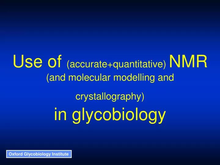
Use of (accurate+quantitative) NMR (and molecular modelling and crystallography) in glycobiology. Oligosaccharides. Formed by linking monosaccharides via a glycosidic linkage. Each monosaccharide can be a or b at the anomeric carbon (position 1)

E N D
Use of (accurate+quantitative) NMR (and molecular modelling and crystallography) in glycobiology
Oligosaccharides Formed by linking monosaccharides via a glycosidic linkage • Each monosaccharide can be a or b at the anomeric carbon (position 1) • Linkages can be made to the 2, 3, 4 or 6 positions (if available) • Several residues can be linked to a single monosaccharide (branching) Much higher degree of complexity than other biopolymers
Features of protein glycosylation • There are several linkage types to proteins, including - • N-linked via Asn (requires an Asn-X-Ser/Thr sequon) • O-linked via Ser or Thr • GPI anchor via peptide C-terminus • Glycosylation is a co-/post-translational modification - • Depends on cell machinery as well as protein sequence • Species, tissue and disease-state specific • A well-defined, reproducible set of oligosaccharides is found at any given site on the protein - • A glycoprotein consists of an ensemble of glycoforms
Oligosaccharide 1D NMR spectrum NeuAc1Gal2Man3GlcNAc4Fuc1
Conformations of oligosaccharides Formed by linking monosaccharides via a glycosidic linkage Monosaccharide rings are rigid and have a well-defined conformation independent of environment The conformational analysis of an oligosaccharide reduces to determining the torsion angles about each glycosidic linkage (2 or 3 torsion angles per residue).
O O O O O O Conformations of saccharide linkages - information required to define linkage structure • For each distinct conformer • Average linkage torsion • angles • Fluctuations around the • average position • For the ensemble of conformers • Relative populations of the • different conformers • Rate of transition between • the conformers
Conformations of saccharide linkages - information available • X-ray crystallography - • Most oligosaccharides and glycoproteins either do not crystallise or give no resolvable electron density for the glycan. Glycans that can be seen are incomplete. • Can give average properties of linkages. • Nuclear Magnetic Resonance Spectroscopy - • Experimental structural parameters (inter-nuclear distances and torsion angles) averaged on a msec timescale. • Can be interpreted in terms of a structure if it is assumed that there is a single well-defined conformation. • Molecular Dynamics Simulations - • Theoretical dynamic structures on a nsec timescale. • Can be interpreted in terms of a structure if it is assumed that the theory is correct.
Applications of crystallographic databases of glycosidic linkages • Estimate range of possible allowed conformations • Check experimental glycan structures • correct “incorrect” structures • identify “distorted” structures • Build “average” glycan structures for X-ray refinement • Provide test data for forcefield validation • Model glycoprotein structures and glycan:protein interactions Structural Assessment of Glycosylation Sites (SAGS) database https://sags.biochim.ro/
Crystal structures containing glycosidic linkages - 2002 Crystal structures containing glycosidic linkages - 2009
Crystallographic refinement problems 1dpj 1wbl PDB structure: 6 Bond angle 80 3 sp2 (planar) carbon Biosynthetic structure: 6 3 incorrect monomers or linkages ring or linkage distortion Useful tool: http://www.dkfz-heidelberg.de/spec/glycosciences.de/tools/pdbcare/ Dr M. Crispin – personal communication
Crystallographic glycosidic linkage conformers - Man 1-4 GlcNAc linkage 250 180 C1-O-C4’-C3’ 200 120 150 100 60 50 Frequency C1-O-C4’-C3’ 0 0 250 O5-C1-O-C4’ 200 -60 150 100 -120 50 0 -180 -180 -120 -60 0 60 120 180 -180 -120 -60 0 60 120 180 O5-C1-O-C4’ Torsion angle Major cluster – 56% of structures
Crystallographic glycosidic linkage conformers - 2009
Crystallographic glycosidic linkage conformers - chitobiose From 1999 to 2009: Number of structures increased by 1,800% Percentage of structures within the major cluster decreased from 90% to 70%
Conformational constraints from NOEs • Presence of an NOE - • gives a value for the average 1/r6 inter-proton distance (on a msec timescale). • Absence of an NOE - • gives a minimum value (of3.5 Å) of the inter-proton distance for all significantly populated conformations.
H1 - H2’ distance O 140 H 90 r 40 O H C1-O-C2’-H2’ -10 -60 O -110 NOE intensity 1/r6 -160 -110 -60 -10 40 90 140 H1-C1-O-C2’ 2.75 - 3.0 Å Conformational constraints from NOEs
1 ROE 0.675 0.5 0.385 0 NOE -1 -1 0.1 1 10 Nuclear Magnetic Resonance Spectroscopy - steady state NOE intensity and mobility Generally assume that c is constant for any given disaccharide. Then, can use an intra residue NOE to calibrate the cross-linkage NOEs. Intensity oc
5.6 5.4 4.0 3.8 3.6 Nuclear Magnetic Resonance Spectroscopy - effects of local correlation times ROESY NOESY Glc1-2Glc Chemical shift (ppm)
Bo Nuclear Magnetic Resonance Spectroscopy - differing local correlation times The local correlation time for a given proton pair depends on the reorientation of the inter-nuclear vector with respect to the applied magnetic field. Anisotropic tumbling - Inter-nuclear vectors will reorient at different rates depending on their angle with respect to the principle axis. Internal flexibility - Inter-nuclear vectors will reorient at different rates depending on the degree of local flexibility. H H H H H H H H
Nuclear Magnetic Resonance Spectroscopy - NOE intensity, distance and motion • Using the two-spin approximation: • rij is the distance between the two nuclei • B is a constant • is the precession frequency • J is the spectral density function given by: • ij is the local correlation time for the pair of nuclei Poveda, et al (1997) Carbohydr. Res., 300, 3-10
Nuclear Magnetic Resonance Spectroscopy - measuring local correlation times o = 500 MHz 3 (ROE) (T-ROE) 2 Ratio of build-up rates 1 (NOE) (ROE) 0 (NOE) (T-ROE) -1 0 0.2 0.4 0.6 c (ns) Poveda, et al (1997) Carbohydr. Res., 300, 3-10
Effective local correlation times measured by (NOE)/(T-ROE) ratio Distances calculated from NOESY data assuming a rigid molecule Distances calculated from NOESY data allowing for local correlation times 200 ps 2.13 Å 2.40 Å 2.30 Å 2.30 Å 280 ps 289 ps 2.30 Å 2.24 Å Local correlation times - Glc3ManOMe Glc1-2Glc o = 500 MHz
H2 H1 H3 H1-H2 NOE NOE intensity H1-H3 NOE 0 0 Mixing time Nuclear Magnetic Resonance Spectroscopy - NOE intensity and mixing time • Short mixing time - • Observed NOE intensity is • linear with mixing time • 1/r6 • Long mixing time - • Observed NOE intensity • non-linear with mixing time • includes spin-diffusion • depends on 3-D structure • and correlation time (dynamics)
Bo 1H 13C Partial alignment in a liquid crystal - Residual Dipolar Coupling 1JCH(aligned) = 1JCH(unaligned) + RDC
Bo Partial alignment of oligosaccharides in a liquid crystal H Sij is assumed to be constant for a given monosaccharide residue. H O H H H O The General Degree of Order (GDO) parameter is a measure of molecular motion of a given residue. H H O Tian, et al (2001) J. Am. Chem. Soc., 123, 485-492
GLYCAM force-field parameters Exo-anomeric effect Partial charges AMBER Woods, et al (1995) J. Phys. Chem., 99, 3832-3846
Molecular Dynamics of Oligosaccharides • Amber Force Field, parameterised to fit ab initio calculations - • Exo-anomeric effect (torsion angle terms) • Solvation (partial charges) • Obtain starting linkage geometries from calculations on disaccharides • Run unconstrained molecular dynamics simulations in water • Compare results to experimental data - • average data from X-ray crystallography • torsion angle constraints from NMR (NOEs and 3JHH) • back calculate NOE build-up curves
Solution conformation of oligomannose N-linked oligosaccharides Dr Rob Woods Dr Chris Edge Dr Andrei Petrescu, Institute of Biochemistry, Bucharest
Oligomannose glycans Dol Complex glycans Hybrid glycans GlcNAc Man Glc Gal Fuc NeuAc N-glycan biosynthesis ER Golgi
Man Man Man Man D3 B 6 3 Man 6 3 4’ Man GlcNAc GlcNAc D2 A 3 2 1 Man Man Man D1 C 4 Man a1-2 Man linkage Schematic structure of Man9GlcNAc2 1,6 arms 1,3 arm Mana1-2Man linkages occur in oligomannose type N-glycans, polysaccharides such as mannan and GPI anchors.
Crystallographic glycosidic linkage conformers - 2009
Crystallographic glycosidic linkage structures - Man a1-2 Man linkage (2009) 180 70 185 structures H1-C1-O-C2’ C1-O-C2’-H2’ 60 120 50 60 40 0 Population C1-O-C2'-H2' 30 -60 20 -120 10 -180 0 -180 -120 -60 0 60 120 180 -180 -120 -60 0 60 120 180 H1-C1-O-C2' Torsion Angle
5.4 4.8 4.4 4.0 3.6 Chemical Shift (ppm) Man9GlcNAc2 NOESY traces 4’:C1H 4’:C2H 3:C6H 3:C6H 4’:C3H HDO 3:C5H A:C1H 4’:C3H A:C5H + D2:C5H A:C2H D2:C1H 4’:C2H A:C3H D2:C1H A:C2H D2:C2H A:C1H A:C3H
Man9GlcNAc2 NMR torsion angle map D1-C Man2Man linkage C1H - C1H’ = 2.80 - 3.15 Å C1H - C2H’ = 2.05 - 2.30 Å C1H - C3H’ = 3.1 - 3.7 Å C5H - C1H’ = 2.4 - 2.9 Å C1H - C4H’ > 3.5 Å
Man9GlcNAc2 Molecular Dynamics D1-C Man2Man linkage H1-C1-O-C2’ C1-O-C2’-H2’
f = -60, y = -60 f = -60, y = +60 OH OH HO OH HO O O HO OH OH HO O OH O OH HO O OH OH OH O O H OH H Hydrogen bond Hydrophobic interactions Conformation of the Man a1-2 Man linkage
Man9GlcNAc2 NMR torsion angle map A-4’ ManMan linkage 140 90 • C1H - C2H’ = 2.85 - 3.20 Å • C1H - C3H’ = 2.00 - 2.25 Å • C1H - C4H’ = 2.75 - 3.10 Å • C5H - C2H’ = 2.55 - 2.85 Å • Individual MD • conformations 40 C1-O-C3’-H3’ -10 -60 -110 -110 -60 -10 40 90 140 H1-C1-O-C3’
Man9GlcNAc2 NMR build up curves A-4’ ManMan linkage 10 8 6 NOE Intensity (%) C1H - C3H’ C5H - C2H’ C1H - C4H’ C1H - C2H’ 4 2 0 0 500 1000 1500 2000 Mixing time (ms)
Schematic structure of Man9GlcNAc2 1,6 arms Man Man Man Man D3 B 6 3 Man 6 3 4’ Man GlcNAc GlcNAc D2 A 3 2 1 Man Man Man D1 C 4 1,3 arm
Molecular Dynamics of Man9GlcNAc2 Torsion angle Simulation time (ps)
Molecular Dynamics of Man9GlcNAc2 Overlay of structures (all atoms) from 1000 ps Side view Top view
Flexibility of the tri-glucosylated cap of immature N-linked glycans Mukram Mackeen Drs Andy Almond and Michael Deschamps, Dept. of Biochem., Oxford Dr Terry Butters Dr Antony Fairbanks, Dept. of Chem., Oxford
Dol P P ER processing and recognition of N-linked glycans - protein folding and quality control Nascent peptide OST Glucosidase I Glucosidase II Glucosidase II Glucosyl transferase Recognised by Calnexin and Calreticulin ER resident chaperones involved in protein folding Golgi ER ERAD
Man Man Man Man D3 B 6 3 Man 6 3 4’ Man GlcNAc GlcNAc D2 A 3 2 1 Glc Glc Glc Man Man Man G1 G2 G3 D1 C 4 Schematic structure of Glc3Man9GlcNAc2 1,6 arms 1,3 arm
Multifunctional roles of GlcxMan9GlcNAc2 Glc3Man Glc 1 Glycan remodelling to produce mature glycans Chaperone assisted folding Calnexin and calreticulin binding OGT substrate Protein folding quality control ERAD recognition Protein N-glycosylation OST substrate binding GlcNAc 1
Crystallographic glycosidic linkage conformers - 2009 Glc3Man linkages: Glca1-2Glc 0 Glca1-3Glc 1 Glca1-3Man 0
-150 -150 -150 -50 -50 -50 +50 +50 +50 +150 +150 +150 Glc3Man NMR torsion angle maps G1-G2 G2-G3 G3-D1 +150 +50 - C1-O-C’x-H’x -50 -150 - H1-C1-O-C’x Solid lines : positive constraints Dotted lines : negative constraints White – H1-H’x-1Grey – H5-H’x-1 Red – H1-H’xPink – H5-H’x Green – H1-H’x+1Yellow – H5-H’x+1
4.9 D1:1 D1:1 A:1 A:1 5.1 G3:1 G3:1 C:1 C:1 4:1 4:1 5.3 2 – 1H (ppM) 4.9 5.1 5.3 116 115 114 113 112 1 – 13C (ppM) Residual dipolar coupling - Glc3Man7GlcNAc2 Unaligned Aligned